Copyright
©2010 Baishideng.
World J Hepatol. May 27, 2010; 2(5): 201-202
Published online May 27, 2010. doi: 10.4254/wjh.v2.i5.201
Published online May 27, 2010. doi: 10.4254/wjh.v2.i5.201
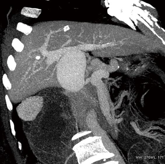
Figure 1 Multi intensity projection shows a large aneurysm (maximum diameter 6.
3 cm) of the MPV that arose 1 cm after the spleno-mesenteric confluence.
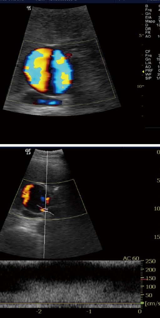
Figure 2 Color-Doppler US showsa turbulent flow inside the aneurysm (red and blue color).
An increase velocity of flow isdetected at the origin of the aneurysm.
- Citation: Francesco FD, Gruttadauria S, Caruso S, Gridelli B. Huge extrahepatic portal vein aneurysm as a late complication of liver transplantation. World J Hepatol 2010; 2(5): 201-202
- URL: https://www.wjgnet.com/1948-5182/full/v2/i5/201.htm
- DOI: https://dx.doi.org/10.4254/wjh.v2.i5.201


