Copyright
©2010 Baishideng.
World J Hepatol. May 27, 2010; 2(5): 192-197
Published online May 27, 2010. doi: 10.4254/wjh.v2.i5.192
Published online May 27, 2010. doi: 10.4254/wjh.v2.i5.192
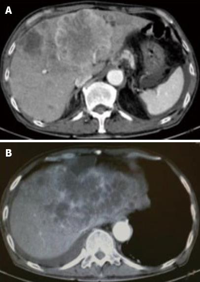
Figure 1 Contrast-enhanced (CE) computed tomography of the abdomen revealed a huge tumor, measuring 11 cm in diameter, which presented with ring enhancement in the arterial phase and central necrosis.
A: In the medial segment; B: Multiple small-sized tumors mainly located in the anterior and medial segments of the liver.
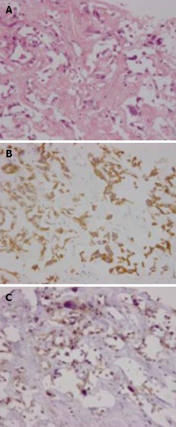
Figure 2 Biopsied specimens from one of the hepatic tumors.
A: A moderately-differentiated adenocarcinoma with wide-spread necrosis, which were not accompanied by hepatic cell components (HE × 200); B: Positive immunostaining for cytokeratin 7 (× 200) in the adenocarcinoma cells; C: Immuno-positive AFP (× 200) in the adenocarcinoma cells.
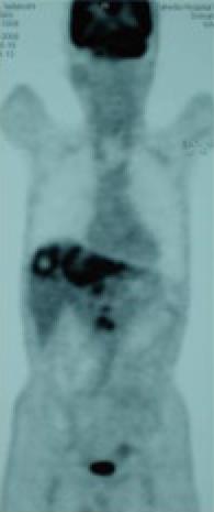
Figure 3 FDG-PET CT after the end of 2 cycles of chemotherapy revealed abnormal uptake in the hepatic tumors in both lobes and the LNs in the liver hilus and para-aorta.
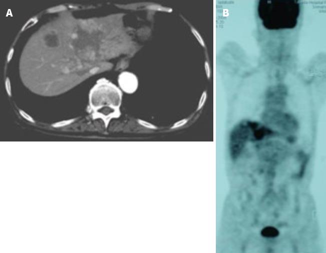
Figure 4 Images after the end of 7 cycles of chemotherapy.
A: CE-CT showed a reduction in the size of a huge tumor seen in the medial segment; B: PET-CT revealed abnormal uptake only in the tumors in the medial segment, and no uptake was observed in tumors from the right lobe and the LNs in the liver hilus and para-aorta.
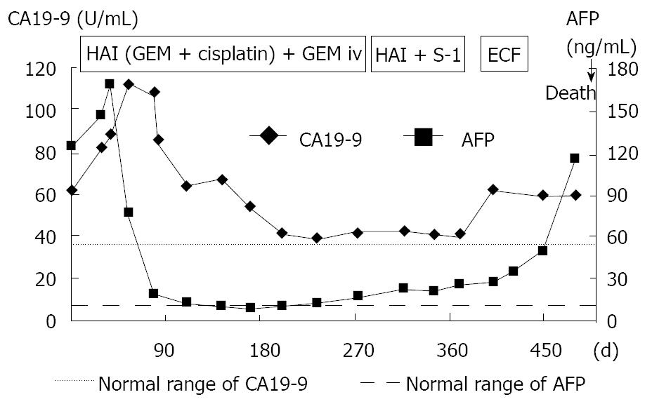
Figure 5 Clinical course and changes in the serum levels of CA19-9 and AFP.
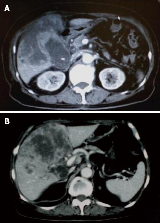
Figure 6 CE-CT after the end of 4 cycles of i.
v. administration chemotherapy showed that the tumor increased in size. A: The GB tumor; B: Liver tumor.
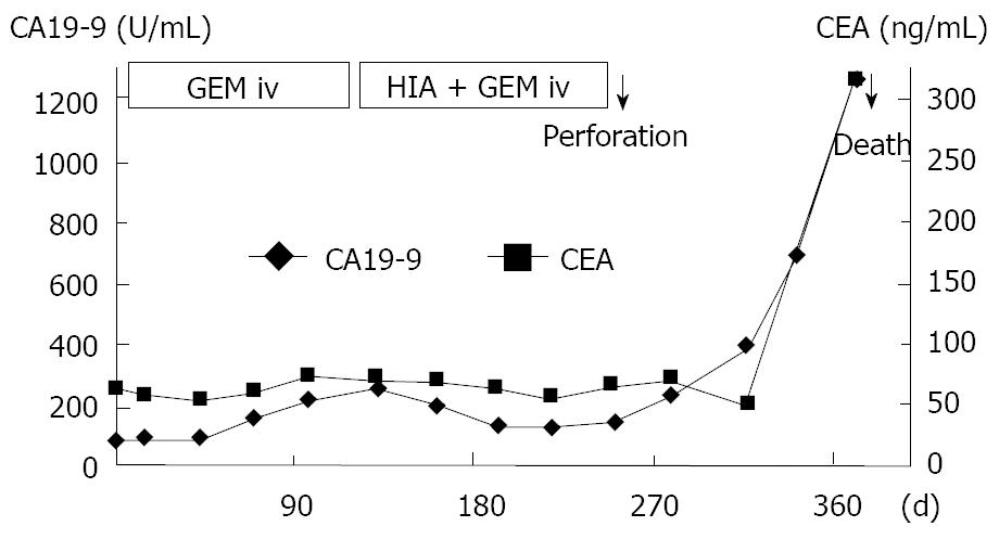
Figure 7 Clinical course and changes in the serum levels of CA19-9 and CEA.
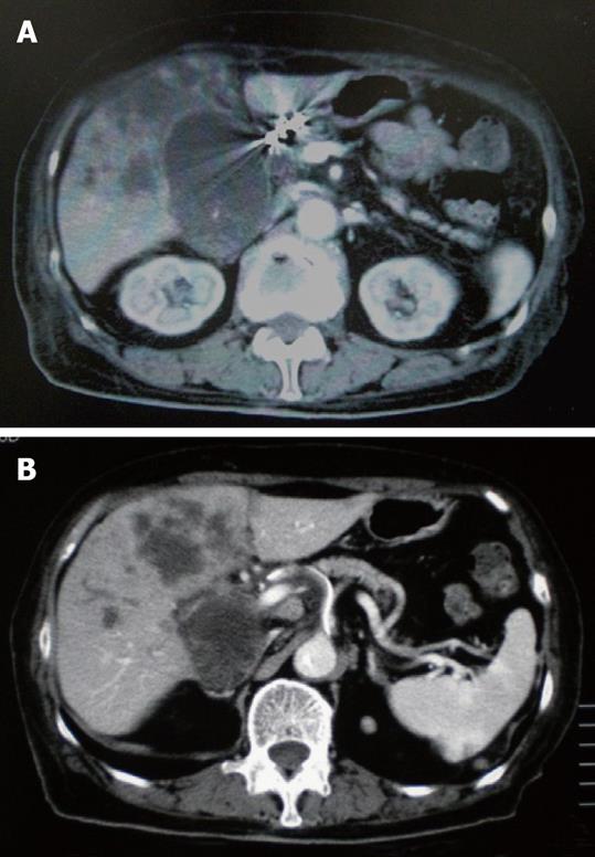
Figure 8 CE-CT after the end of 3 cycles of HAI chemotherapy showed a decrease in the size of tumor.
A: The GB tumor; B: Liver tumor.
- Citation: Nishimura M. A successful treatment by hepatic arterial infusion therapy for advanced, unresectable biliary tract cancer. World J Hepatol 2010; 2(5): 192-197
- URL: https://www.wjgnet.com/1948-5182/full/v2/i5/192.htm
- DOI: https://dx.doi.org/10.4254/wjh.v2.i5.192


