Copyright
©2010 Baishideng.
World J Hepatol. May 27, 2010; 2(5): 185-191
Published online May 27, 2010. doi: 10.4254/wjh.v2.i5.185
Published online May 27, 2010. doi: 10.4254/wjh.v2.i5.185
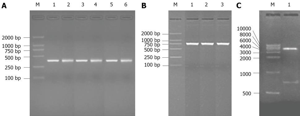
Figure 1 Agarose gel electrophoresis of PCR products.
A: PCR products of VH and VL of scFv-3, scFv-4 and scFv-5. M: DNA marker DL2000; Lane 1: PCR products of the VH gene of scFv-3; Lane 2: PCR products of the VL gene of scFv-3. Lane 3: PCR products of the VH gene of scFv-4; Lane 4: PCR products of the VL gene of scFv-4. Lane 5: PCR products of the VH gene of scFv-5; Lane 6: PCR products of the VL gene of scFv-5; B: PCR products of scFv-3, scFv-4 and scFv-5. M: DNA marker DL2000; Lane 1: PCR products of the scFv-3 gene; Lane 2: PCR products of the scFv-4 gene; Lane 3: PCR products of the scFv-5 gene; C: Digested pCANAB5E vector: M: DNA marker; Lane 1: Digested pCANAB5E vector.
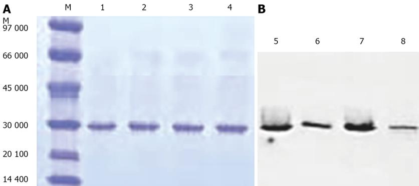
Figure 2 SDS-PAGE and Western blot.
A: SDS gel electrophoresis of affinity-purified scFv fragments under reducing conditions. M: Low molecular weight markers ( kDa); Lane 1: scFv-3; Lane 2: scFv-4; Lane 3: scFv-5; Lane 4: parental scFV HBM; B: Western blot. Lane 5: scFv-3; Lane 6: scFv-4; Lane 7: scFv-5; Lane 8: parental scFV HBM.
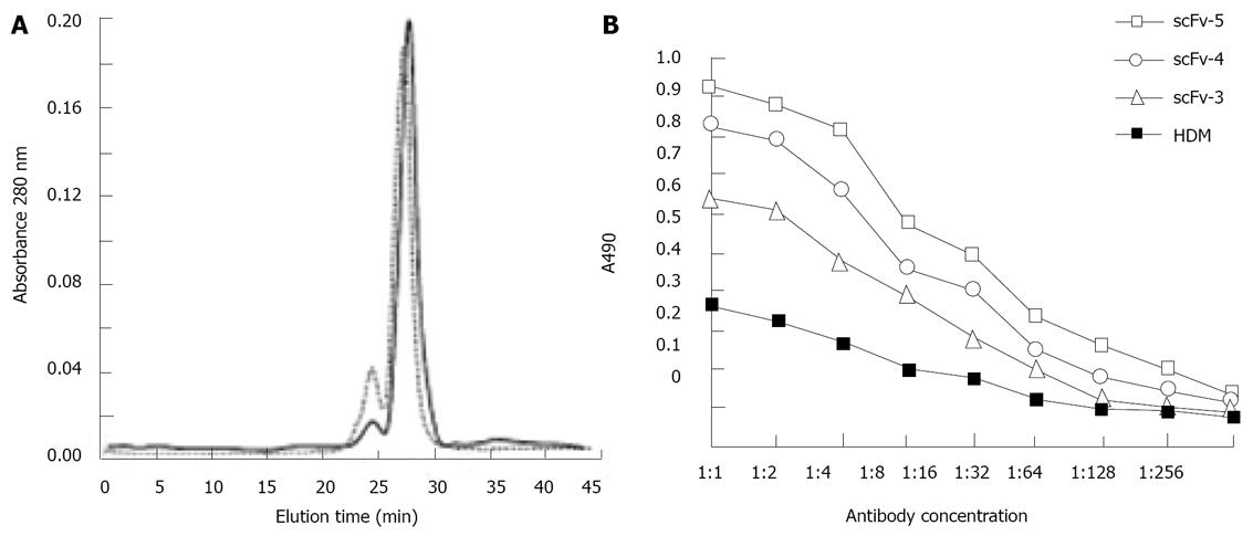
Figure 3 Analysis of soluble scFv dimers.
A: Size exclusion chromatography of affinity purified scFv-3, scFv-4, scFv-5. Superimposed are elution profiles from a Superdex 200 gel filtration column of scFv-4 (solid line) and scFv-3 (dashed line). Elution peaks and molecular weight of calibration reference proteins are Gel Filtration Standard proteins (Biorad); B: Equilibrium binding curves of scFv dimers. Binding activity to hepatocellular carcinoma cell lines: ScFv-5 > scFv-4 > scFv-3 > HDM, with the tendency of decreasing binding activity going by diluted concentration. Determined By Model 680 Microplate Reader (Biorad).
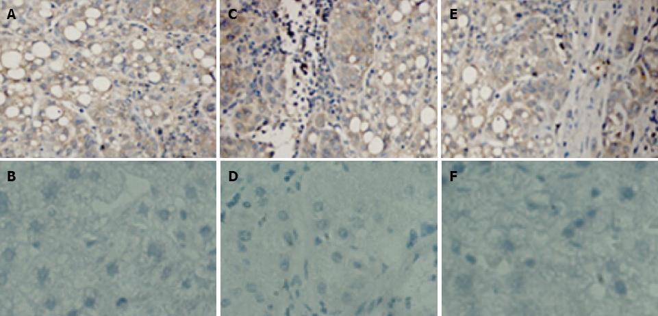
Figure 4 Immunohistochemistry of human hepatocellular carcinoma tissue.
A: Reaction of scFv-3 dimer and human hepatocellular carcinoma tissue; B: Reaction of scFv-3 dimer and non- hepatocellular carcinoma tissue; C: Reaction of scFv-4 dimer and human hepatocellular carcinoma tissue; D: Reaction of scFv-4 dimer and non- hepatocellular carcinoma tissue; E: Reaction of scFv-5 dimer and human hepatocellular carcinoma tissue; F: Reaction of scFv-5 dimer and non- hepatocellular carcinoma tissue. Bound scFv dimers were detected with an antibody directed to the hexahistidine tag (Dianova) and peroxidase-conjugated rabbit anti-mouse immunoglobulins (Dako). Staining was performed with diaminobenzidine (DAB)/H2O2; nuclei were counterstained with hematoxylin. As expected, scFv dimers react with human hepatocellular carcinoma tissue but not with non- hepatocellular carcinoma tissue. The image was captured at 20 × magnification.
- Citation: Bie CQ, Yang DH, Liang XJ, Tang SH. Construction of non-covalent single-chain Fv dimers for hepatocellular carcinoma and their biological functions. World J Hepatol 2010; 2(5): 185-191
- URL: https://www.wjgnet.com/1948-5182/full/v2/i5/185.htm
- DOI: https://dx.doi.org/10.4254/wjh.v2.i5.185


