Copyright
©2010 Baishideng.
World J Hepatol. May 27, 2010; 2(5): 175-179
Published online May 27, 2010. doi: 10.4254/wjh.v2.i5.175
Published online May 27, 2010. doi: 10.4254/wjh.v2.i5.175
Figure 1 Macroscopic and histological aspect of HCC in BALB/c-Abcb4-/--mice.
A: Representative macroscopic view of BALB/c-Abcb4-/--mouse liver at one year of age. Typically, one prominent tumor and a number of smaller tumors are visible; B: Hematoxylin and Eosin (H&E) stained liver section of the invasive tumor front (tumor on the upper right, fibrotic parenchyma on the lower left side, dashed line: HCC tumor capsule, bar represents 100 μm); C: Fifty-fold enlarged Sirius red (SR) stained liver section photographed under polarized light (PL, bar represents 400 μm). Fibrillar collagens appear in red. The tumor capsule is rich in collagen, whereas tumor stroma appears collagen-free.
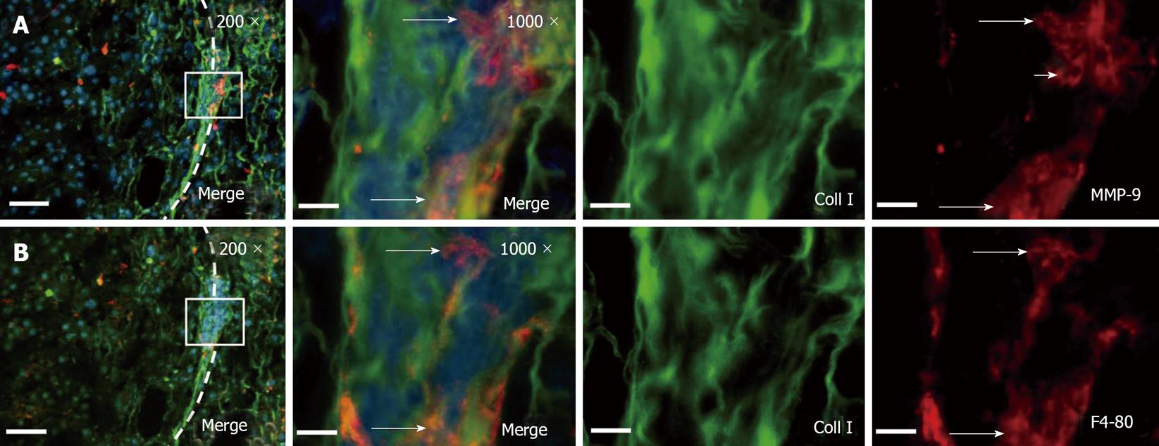
Figure 2 Conglomerates of tumor associated macrophages express MMP-9 within the HCC tumor capsule.
A: Immunofluorescence co-staining of collagen type I (green) and MMP-9 (red) displayed clusters of MMP-9 expressing cells in the tumor capsule. B: After photobleaching of the red fluorescence dye, macrophage marker F4/80 was visualized. The long arrows indicate MMP-9 positive macrophages. The shot arrow indicates an MMP-9 expressing cell which is negative for F4/80. DAPI was used for nucleus staining. The left panels provide a section overview with 200-fold magnification and bars represent 100 μm (left side of the panel: Tumor; dashed line: Tumor capsule; box: This is enlarged to 1000 × magnification in the corresponding panel, bars represent 20 μm). Original green and red channel micrographs (1000 ×) were shown. Images were derived by conventional wide field fluorescence microscopy. Representative digital overlays are shown.
- Citation: Roderfeld M, Rath T, Lammert F, Dierkes C, Graf J, Roeb E. Innovative immunohistochemistry identifies MMP-9 expressing macrophages at the invasive front of murine HCC. World J Hepatol 2010; 2(5): 175-179
- URL: https://www.wjgnet.com/1948-5182/full/v2/i5/175.htm
- DOI: https://dx.doi.org/10.4254/wjh.v2.i5.175


