Copyright
©2010 Baishideng Publishing Group Co.
World J Hepatol. Nov 27, 2010; 2(11): 406-409
Published online Nov 27, 2010. doi: 10.4254/wjh.v2.i11.406
Published online Nov 27, 2010. doi: 10.4254/wjh.v2.i11.406
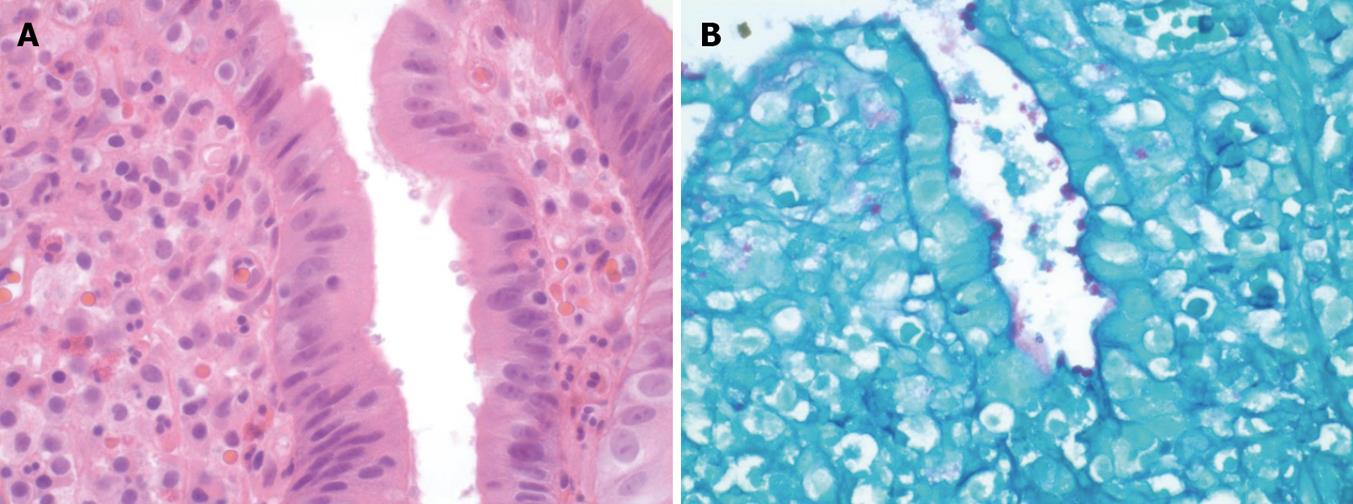
Figure 1 Cryptosporidium microorganisms shown by the biopsy.
A: On the surface epithelium of the terminal ileum; B: On the surface epithelium of the duodenum (Periodic acid schiff stain).
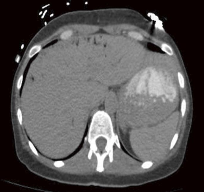
Figure 2 Non-contrast axial computed tomography of the upper abdomen demonstrates multiple peripheral linear branching air density structures consistent with portal venous air.
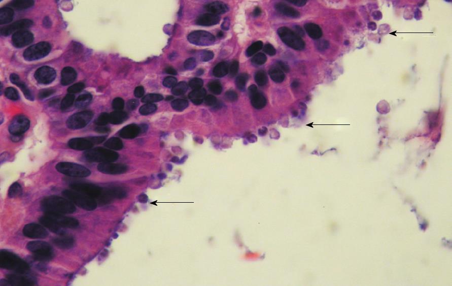
Figure 3 Gallbladder showing Cryptosporidium located on the surface of the epithelium.
- Citation: Lodhia N, Ali A, Bessoff J. Hepatic portal venous gas due to cryptosporidiosis in a patient with acquired immunodeficiency syndrome. World J Hepatol 2010; 2(11): 406-409
- URL: https://www.wjgnet.com/1948-5182/full/v2/i11/406.htm
- DOI: https://dx.doi.org/10.4254/wjh.v2.i11.406


