Copyright
©The Author(s) 2022.
World J Hepatol. Jul 27, 2022; 14(7): 1459-1469
Published online Jul 27, 2022. doi: 10.4254/wjh.v14.i7.1459
Published online Jul 27, 2022. doi: 10.4254/wjh.v14.i7.1459
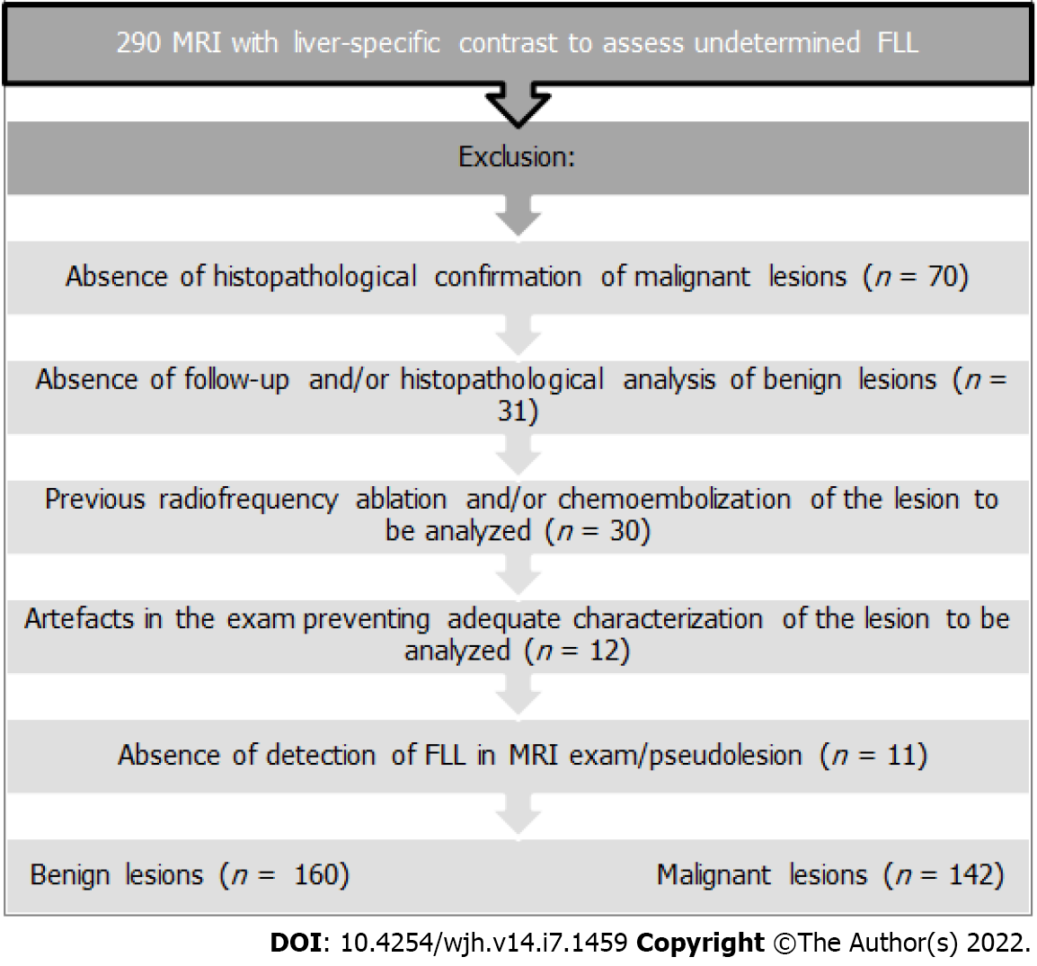
Figure 1 Flowchart: Exclusion criteria.
MRI: Magnetic resonance imaging; FLL: Focal liver lesions.
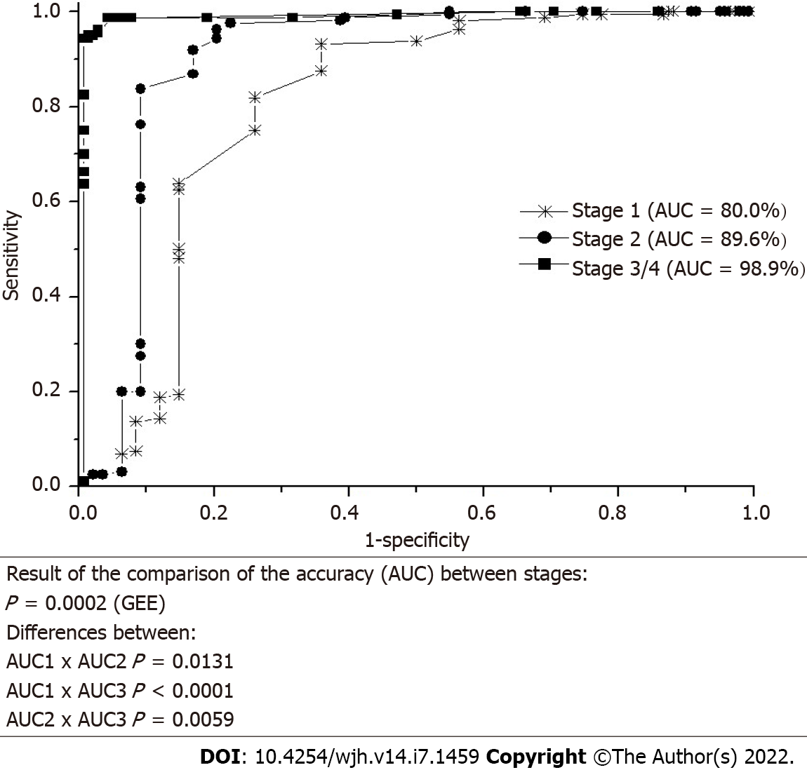
Figure 2 Receiver operating characteristic curves of each stage in relation to the definitive diagnosis and comparison of the accuracy between stages.
AUC: Area under the curve; GEE: Generalized estimating equations.
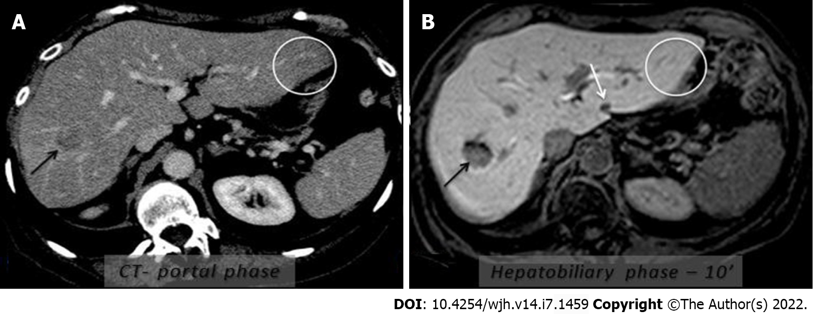
Figure 3 Use of hepatobiliary phases: Detection of small metastasis in a potential patient undergoing liver surgery.
Computed tomography (CT) (A) and magnetic resonance imaging (B) (10-min hepatobiliary phase) showed metastatic lesion in the right lobe (arrow). However, the doubtful/suspected nodule for metastasis on CT was not confirmed in the hepatobiliary phase (isosignal-white circle, pseudolesion). However, another hyposignal nodule (white arrow) in the left lobe was well evidenced in the hepatobiliary phase (it had not been identified on CT), compatible with secondary involvement.
- Citation: Fernandes DA, Dal Lago EA, Oliver FA, Loureiro BMC, Martins DL, Penachim TJ, Barros RHO, Araújo Filho JAB, Eloy da Costa LB, da Silva ÁMO, de Ataíde EC, Boin IFSF, Caserta NMG. Hepatobiliary phases in magnetic resonance imaging using liver-specific contrast for focal lesions in clinical practice. World J Hepatol 2022; 14(7): 1459-1469
- URL: https://www.wjgnet.com/1948-5182/full/v14/i7/1459.htm
- DOI: https://dx.doi.org/10.4254/wjh.v14.i7.1459


