Copyright
©The Author(s) 2021.
World J Hepatol. Jan 27, 2022; 14(1): 260-273
Published online Jan 27, 2022. doi: 10.4254/wjh.v14.i1.260
Published online Jan 27, 2022. doi: 10.4254/wjh.v14.i1.260
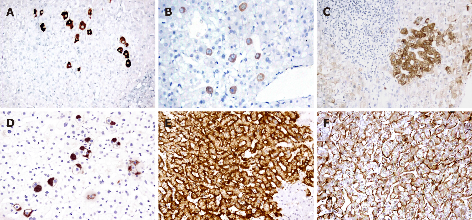
Figure 1 Different staining patterns of hepatitis B surface antigen in liver tissues.
A: Dense cytoplasmic staining in many discrete hepatocytes; B: Diffuse but faint cytoplasmic staining in discrete hepatocytes; C: Diffuse faint cytoplasmic staining in a group of hepatocytes near an inflamed portal area; D: Globular and spotty staining in discrete and in a small group of hepatocytes; E: Submembranous staining in a large group of cells; F: Membranous expression of hepatitis B surface antigen. Original magnifications A and C × 200, B, D, E and F × 400, counter-stained with Mayer’s hematoxylin.
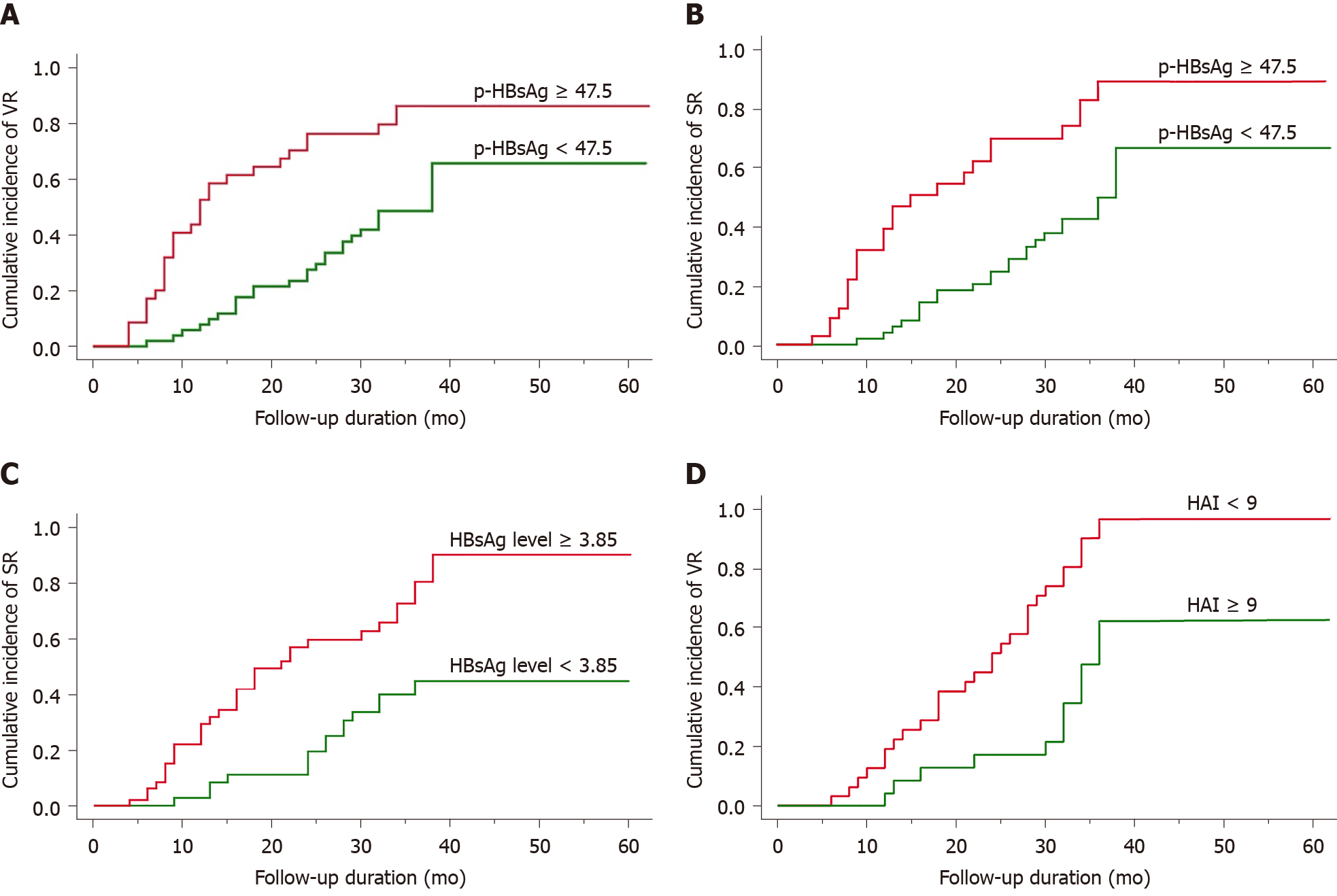
Figure 2 Kaplan-Meier analysis in hepatitis B surface antigen positive and hepatitis B e antigen negative cases (P < 0.
01). A: Cumulative incidence of viral response in hepatitis B e antigen (HBeAg) positive patients is more frequently observed in cases with higher percentage of immunohistochemical hepatitis B surface antigen (p-HBsAg) expression; B-D: In the same group, serological response is also significantly related to p-HBsAg expression (B) and HBsAg levels (C). However, in HBeAg negative in patients, VR is closely related to hepatitis activity index (D). p-HBsAg: Percentage of immunohistochemical hepatitis B surface antigen; HBsAg: Hepatitis B surface antigen; HAI: Hepatitis activity index; VR: Viral response; SR: Serological response.
- Citation: Alpsoy A, Adanir H, Bayramoglu Z, Elpek GO. Correlation of hepatitis B surface antigen expression with clinicopathological and biochemical parameters in liver biopsies: A comprehensive study. World J Hepatol 2022; 14(1): 260-273
- URL: https://www.wjgnet.com/1948-5182/full/v14/i1/260.htm
- DOI: https://dx.doi.org/10.4254/wjh.v14.i1.260


