Copyright
©The Author(s) 2022.
World J Hepatol. Jan 27, 2022; 14(1): 140-157
Published online Jan 27, 2022. doi: 10.4254/wjh.v14.i1.140
Published online Jan 27, 2022. doi: 10.4254/wjh.v14.i1.140
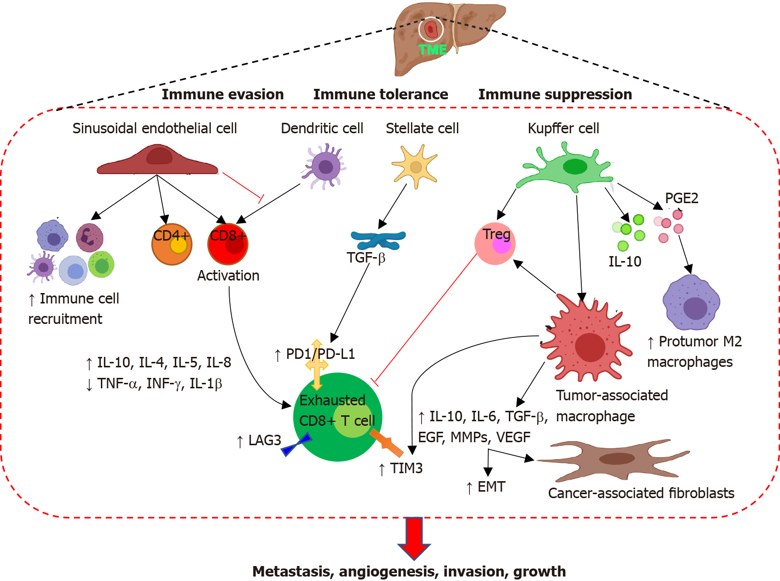
Figure 1 Tumor microenvironment in hepatocellular carcinoma.
Tumor microenvironment comprising of liver sinusoidal endothelial cells, liver resident macrophages or Kupffer cells, hepatic stellate cells, hepatic dendritic cells, tumor-associated macrophages, cytokines, fibroblasts, infiltrating immune cells, pro-tumor M2 macrophages, and growth factors mediate immune suppression, immune tolerance, and immune evasion causing increased tumorigenicity with enhanced evasion, angiogenesis, metastasis, tumor growth. IL: Interleukin; TNF-α: Tumor necrosis factor-alpha; TGF-β: Transforming growth factor-beta; IFN-γ: Interferon-gamma; MMPs: Matrix metalloproteinases; PD-1: Programmed cell death protein 1; PD-L1: Programmed death-ligand 1; PGE2: Prostaglandin E2; EGF: Epidermal growth factor; VEGF: Vascular endothelial growth factor; TIM3: T-cell immunoglobulin and mucin-domain-containing molecule-3; LAG3: Lymphocyte-activation gene 3; EMT: Epithelial mesenchymal transition.
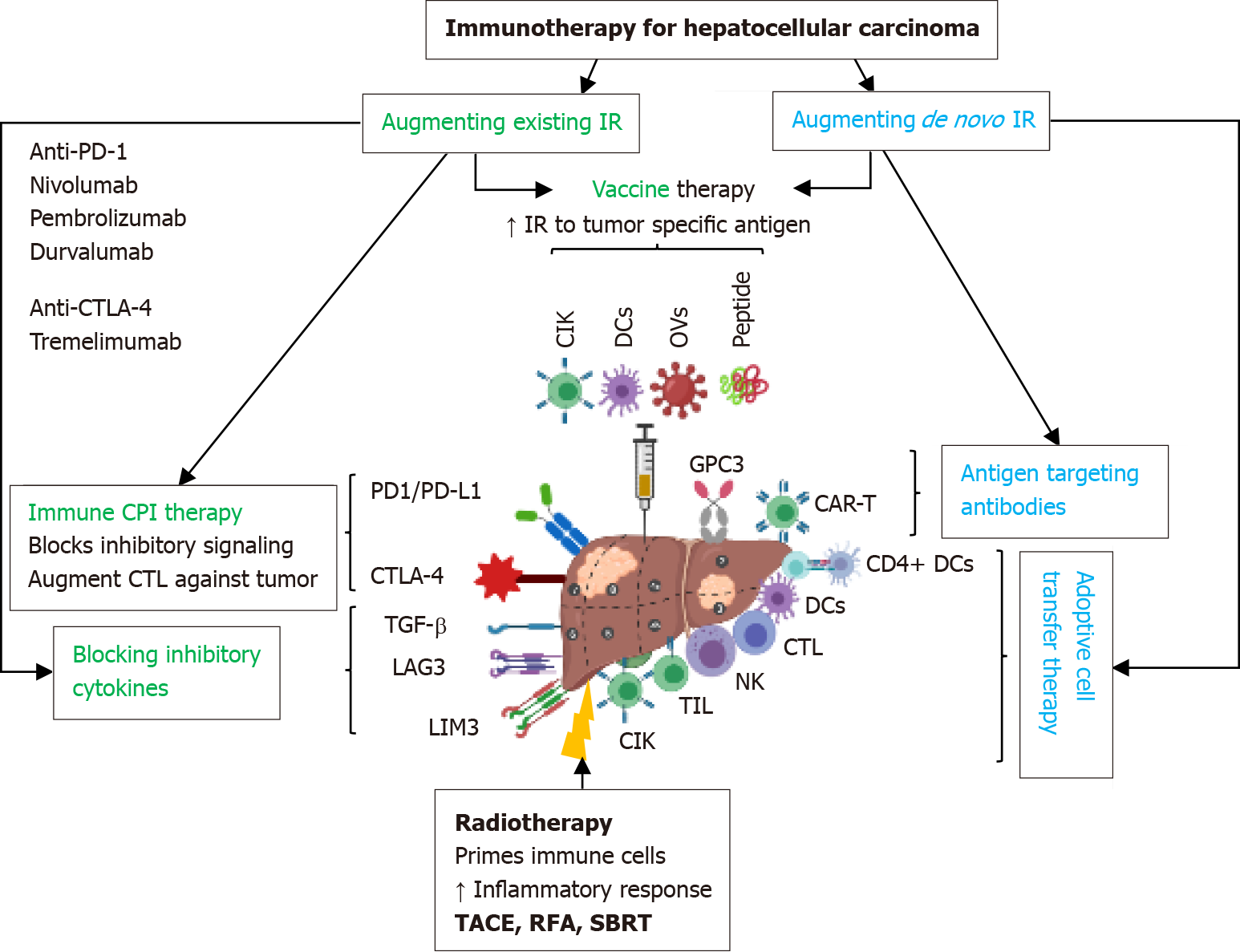
Figure 2 Immunotherapy in hepatocellular carcinoma: Potential strategies and therapeutic targets.
IR: Immune response; PD-1: Programmed cell death protein 1; PD-L1: Programmed death-ligand 1; CTLA-4: Cytotoxic T-lymphocyte associated antigen 4; TGF-β: Transforming growth factor-beta; LAG3: Lymphocyte-activation gene 3; CIK: Cytokine-induced killer; TIL: Tumor-infiltrating lymphocyte; NK: Natural killer; DC: Dendritic cell; GPC3: Glypican-3.
- Citation: Rai V, Mukherjee S. Targets of immunotherapy for hepatocellular carcinoma: An update. World J Hepatol 2022; 14(1): 140-157
- URL: https://www.wjgnet.com/1948-5182/full/v14/i1/140.htm
- DOI: https://dx.doi.org/10.4254/wjh.v14.i1.140


