Copyright
©The Author(s) 2021.
World J Hepatol. Aug 27, 2021; 13(8): 896-903
Published online Aug 27, 2021. doi: 10.4254/wjh.v13.i8.896
Published online Aug 27, 2021. doi: 10.4254/wjh.v13.i8.896
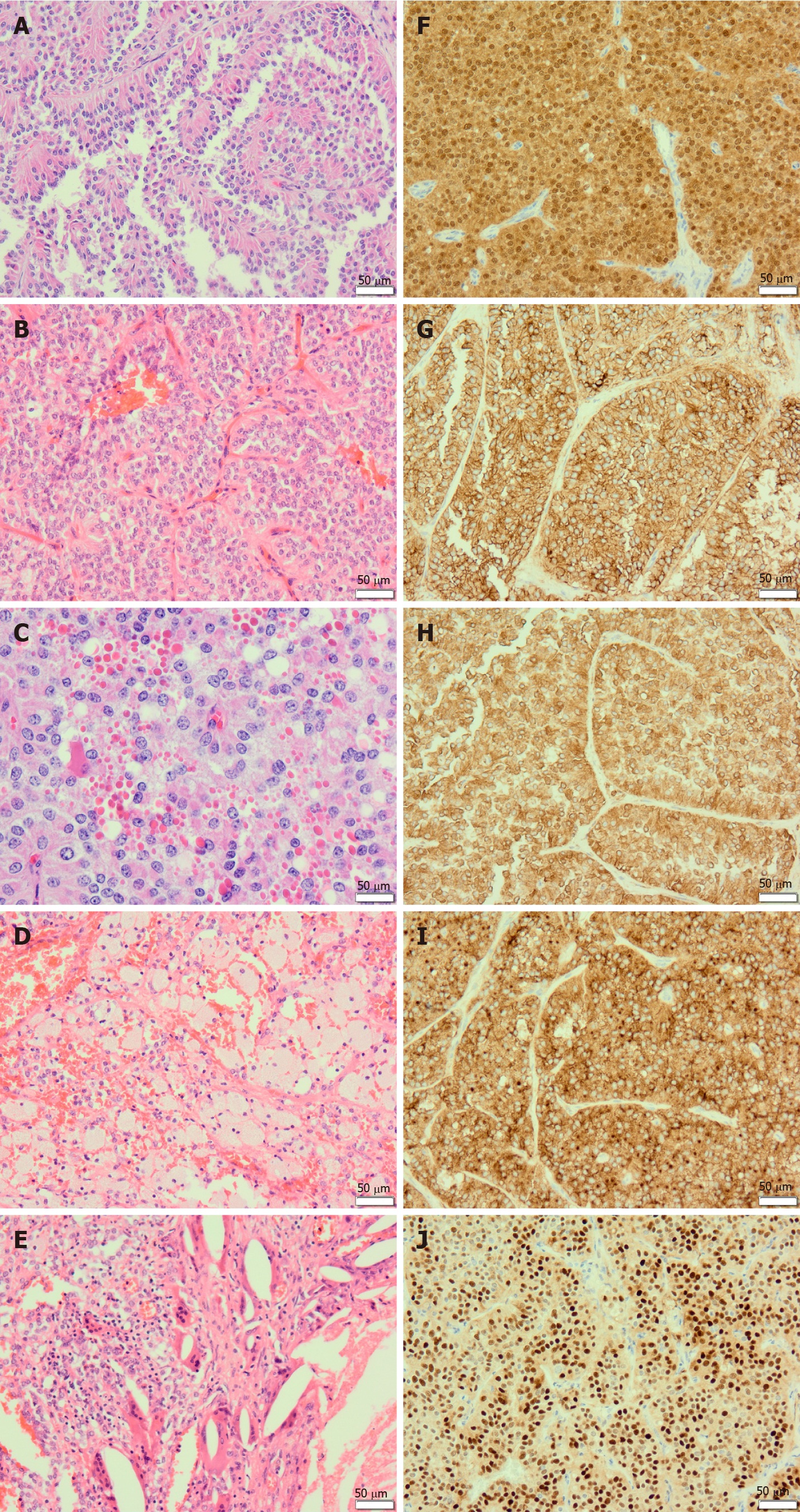
Figure 1 Solid pseudopapillary neoplasm of the pancreas.
A: The tumour shows pseudopapillae formed by poorly cohesive cells arranged around hyalinized fibrovascular stalks (200 ×); B: The tumour consists of cells forming solid and pseudopapillary structures (200 ×); C: Solid pseudopapillary neoplasm of the pancreas shows characteristic eosinophilic cytoplasmic hyaline globules (400 ×); D: An example showing foamy histiocytes (200 ×); E: These are cholesterol clefts surrounded by foreign body giant cells (200 ×); F: The tumour shows nuclear and cytoplasmic expression of β-catenin (200 ×); G: The tumour shows immunolabelling for CD56 (200 ×); H: The tumour is positive for vimentin (200 ×); I: The tumour shows immunolabelling for CD10 (200 ×); J: Solid pseudopapillary neoplasm of the pancreas shows nuclear positivity for cyclin D1 (200 ×).
- Citation: Omiyale AO. Solid pseudopapillary neoplasm of the pancreas. World J Hepatol 2021; 13(8): 896-903
- URL: https://www.wjgnet.com/1948-5182/full/v13/i8/896.htm
- DOI: https://dx.doi.org/10.4254/wjh.v13.i8.896


