Copyright
©The Author(s) 2021.
World J Hepatol. Aug 27, 2021; 13(8): 868-878
Published online Aug 27, 2021. doi: 10.4254/wjh.v13.i8.868
Published online Aug 27, 2021. doi: 10.4254/wjh.v13.i8.868
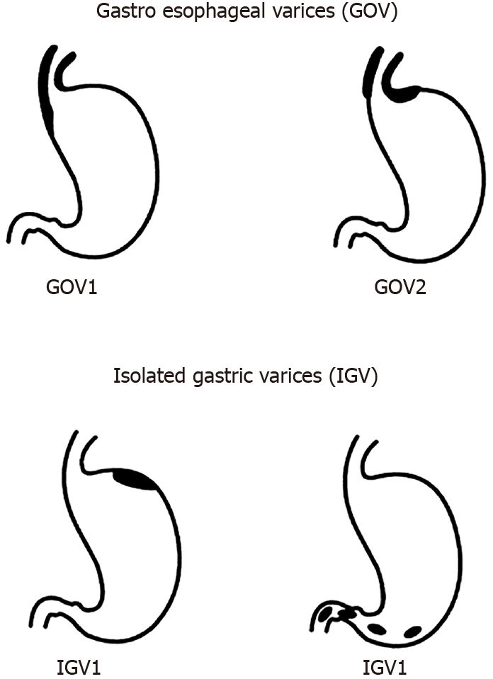
Figure 1 Classification of gastric varices according to Sarin et al[2].
Citation: Sarin SK, Lahoti D, Saxena SP, Murthy NS, Makwana UK. Prevalence, classification and natural history of gastric varices: A long-term follow-up study in 568 portal hypertension patients. Hepatology 1992; 16: 1343-1349. Copyright © The Authors 1992. Published by John Wiley and Sons.
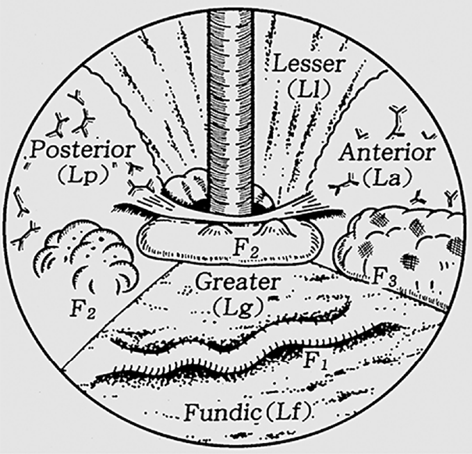
Figure 2 Classification of gastric varices according to Hashizume et al[9].
Citation: Hashizume M, Kitano S, Yamaga H, Koyanagi N, Sugimachi K. Endoscopic classification of gastric varices. Gastrointestinal Endoscopy 1990; 36: 276-280. Copyright © The Authors 1990. Published by Elsevier.
Figure 3 Endoscopic variceal banding of gastroesophageal varices 1.
A: Pre-banding forward view of varix; B: Pre-banding retroflexed view of varix; C: Immediately post-banding of varix.
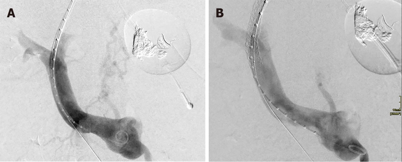
Figure 4 Transjugular intrahepatic portosystemic shunt decompressing gastric varix.
A: Pre-transjugular intrahepatic portosystemic shunt (TIPS) with contrast toward gastric variceal bed; B: Post-TIPS with no contrast flow toward gastric variceal bed.
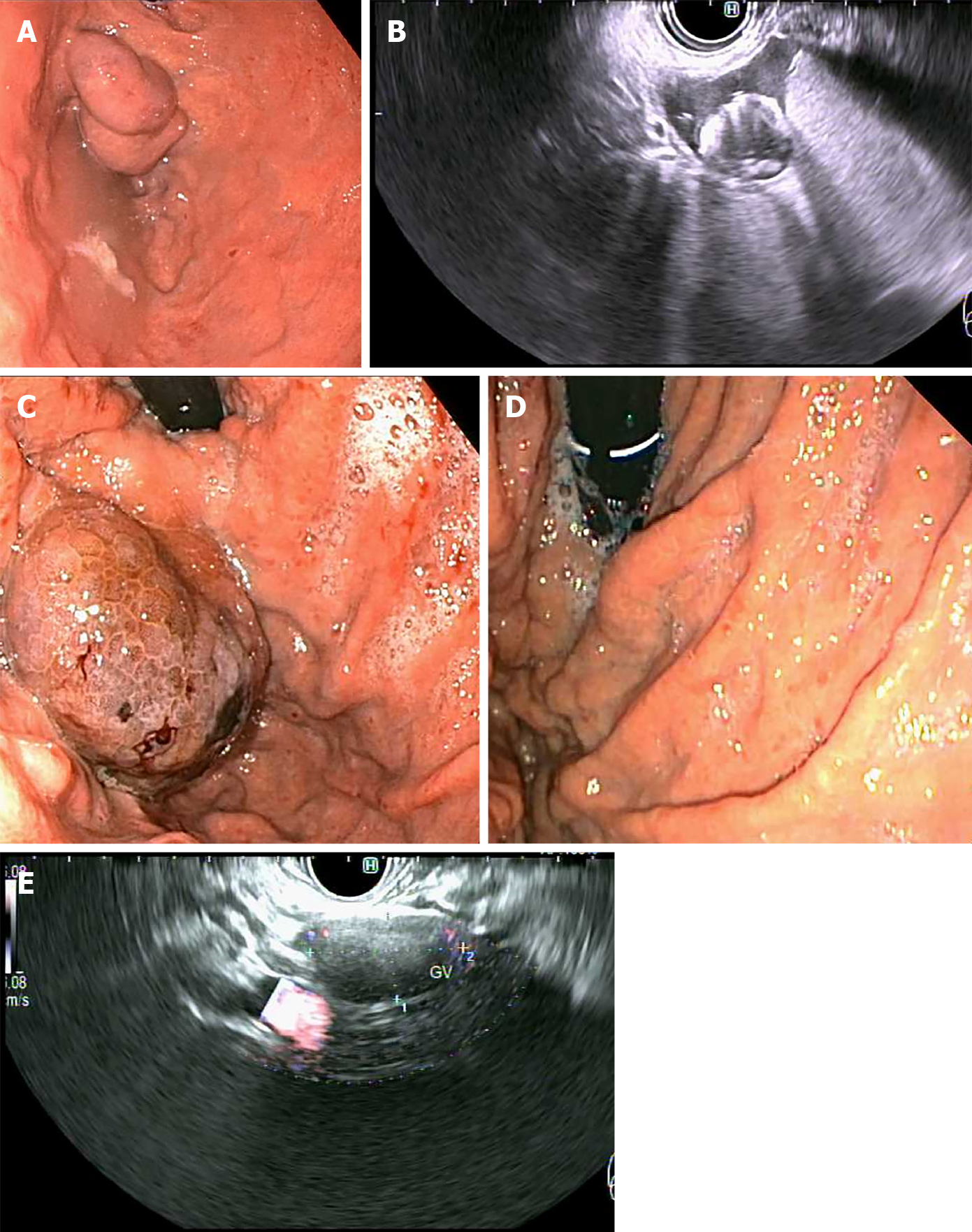
Figure 5 Primary prophylaxis of isolated gastric varices 1 with five 0.
18 10 mm micronester coils in combination 1 mL cyanoacrylate glue. A: Pre-treatment endoscopic appearance; B: Endoscopic ultrasound (EUS) appearance immediately post-deployment of coils; C: Endoscopic appearance 1-mo post-treatment; D: Complete obliteration 4-mo post-treatment; E: EUS appearance of varix with no Doppler flow 4-mo post-treatment.
- Citation: Vaz K, Efthymiou M, Vaughan R, Testro AG, Lew HB, Pu LZCT, Chandran S. Unpacking the challenge of gastric varices: A review on indication, timing and modality of therapy. World J Hepatol 2021; 13(8): 868-878
- URL: https://www.wjgnet.com/1948-5182/full/v13/i8/868.htm
- DOI: https://dx.doi.org/10.4254/wjh.v13.i8.868


