Copyright
©The Author(s) 2021.
World J Hepatol. May 27, 2021; 13(5): 611-619
Published online May 27, 2021. doi: 10.4254/wjh.v13.i5.611
Published online May 27, 2021. doi: 10.4254/wjh.v13.i5.611
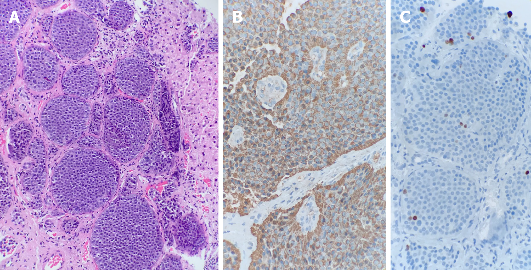
Figure 1 Anatomic pathology report of the tumor biopsy.
A: Hematoxylin and eosin staining showing neoplastic neuroendocrine cells with a typical insular pattern infiltrating hepatic parenchyma; B: Chromogranin A positivity with granular/dot-like cytoplasmic staining; C: Ki-67 staining shows a proliferation rate of 1. 26% in the hot-spot, and the neoplasia was graded as G1.
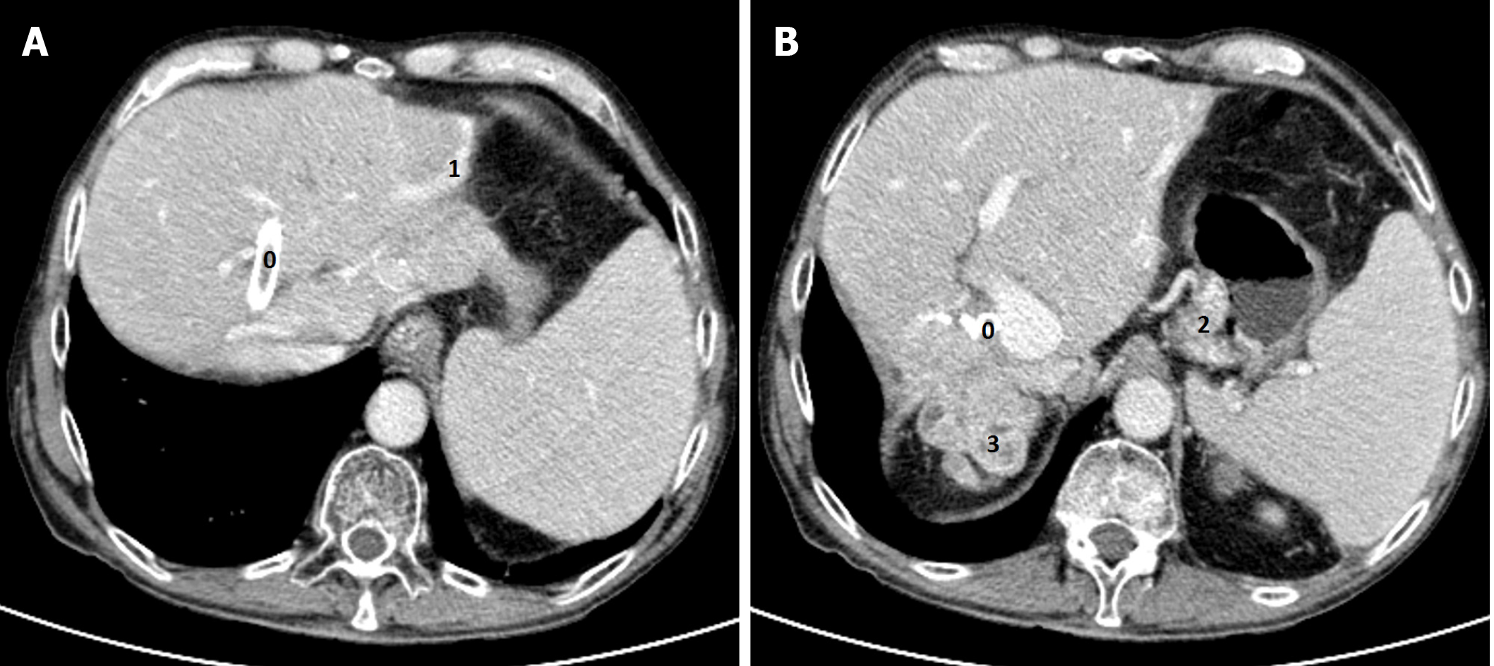
Figure 2 Contrast-enhanced computed tomography (portal phase).
A: Biliary prosthesis (0), and abnormal vascular hepatic vein in the most marginal aspect of the left liver lobe related to a portosystemic shunt (1); B: Enlarged vein in the gastrohepatic ligament associated with small gastric mural varicose veins (2). Metastatic lesions at the hepatic hilum (3).
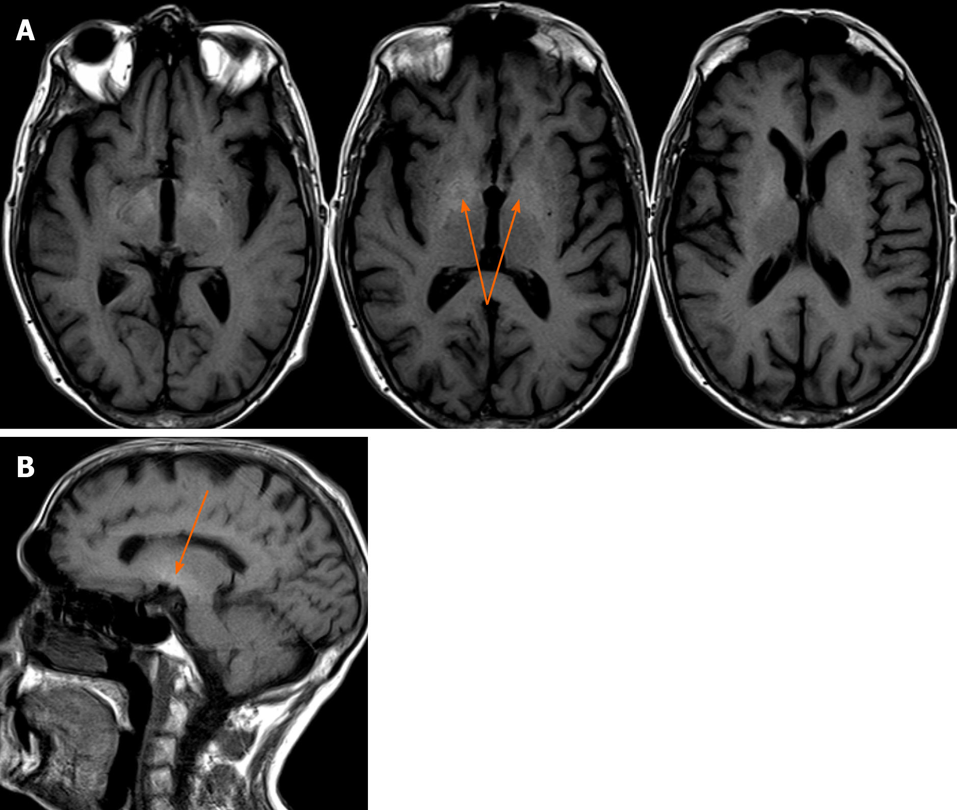
Figure 3 Brain magnetic resonance imaging.
A: Axial; B: Sagittal projection showing T1-weighted imaging, hyperintense signal (arrow) within a lentiform nucleus extending into the midbrain.
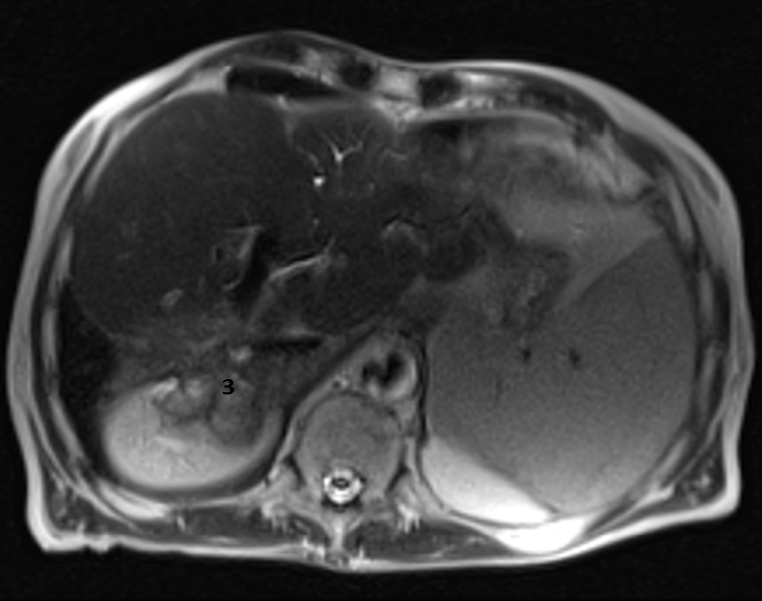
Figure 4 Liver magnetic resonance imaging.
Axial T2-weighted imaging HASTE magnetic resonance imaging. Multiple irregular right liver metastatic lesions (3).
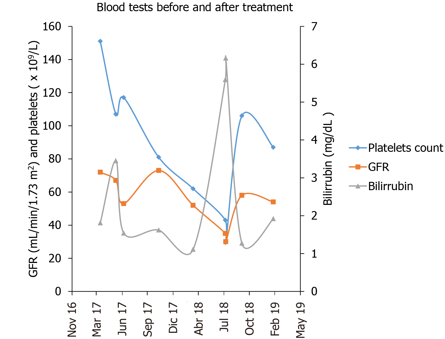
Figure 5 Blood tests before and after treatment.
Laboratory test parameters showing platelet count (× 109/L), glomerular filtration rate (GFR) (in mL/min/173 m2) and total bilirubin (mg/dL) before and after peptide-receptor radionuclide therapy treatment.
- Citation: Mirallas O, Saoudi N, Gómez-Puerto D, Riveiro-Barciela M, Merino X, Auger C, Landolfi S, Blanco L, Garcia-Burillo A, Molero X, Salcedo-Allende MT, Capdevila J. Acquired hepatocerebral degeneration in a metastatic neuroendocrine tumor long-term survivor — an update on neuroendocrine neoplasm’s treatment: A case report. World J Hepatol 2021; 13(5): 611-619
- URL: https://www.wjgnet.com/1948-5182/full/v13/i5/611.htm
- DOI: https://dx.doi.org/10.4254/wjh.v13.i5.611


