Copyright
©The Author(s) 2021.
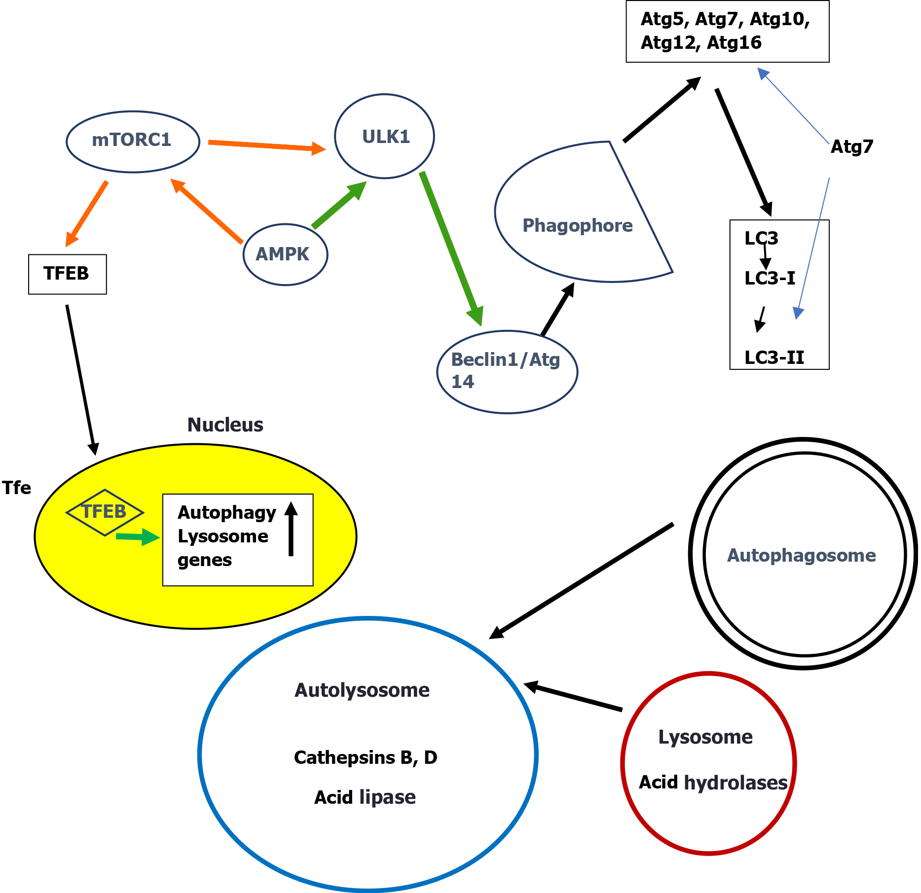
Figure 1 A simplified scheme of the macroautophagy pathways in the liver.
Initiation starts with activation of the unc-51-like kinase 1 complex (ULK1, Atg1 in yeast) followed by beclin 1(Atg6 in yeast) and a subsequent cascade of Atg proteins leading to autophagosome formation where LC3 (Atg8 in yeast) is implicated. LC3 is further processed to form initially LC3-I and then LC3-II. Fusion of the autophagosomes with lysosomes form the autolysosome where acid proteases (among which cathepsins are important) and lipases degrade proteins and lipids. Initiation of autophagy is controlled by two metabolic sensors the mammalian target of rapamycin complex 1 (mTORC1) and the AMP-activated protein kinase (AMPK). mTORC1 negatively regulates autophagy inhibiting ULK1. AMPK suppresses mTORC1 activity. The long-term regulation of autophagy is carried out by transcription factor EB (TFEB), the main regulator of lysosomal biogenesis and autophagy. Under nutrient-rich conditions, mTORC1 phosphorylates TFEB and retains TFEB in the cytosol. Orange arrows: Inhibition. Green arrows: Positive regulation. For details see Ref.[21,29,31]. mTORC1: Mammalian target of rapamycin complex 1; TFEB: Transcription factor EB; ULK1: Unc-51-like kinase 1 complex.
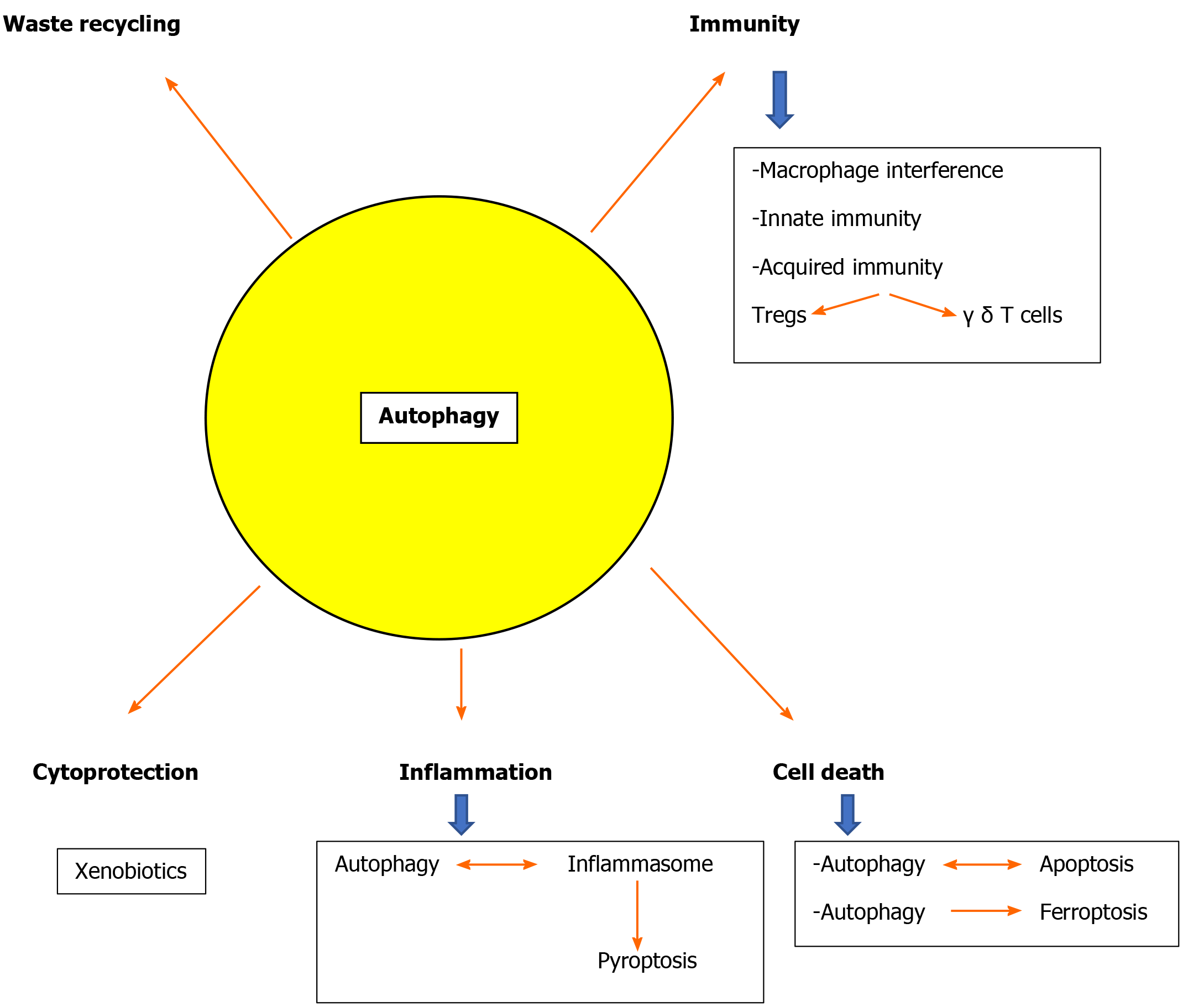
Figure 2 Implications of autophagy in critical cellular functions in the liver.
For details see text.
- Citation: Kouroumalis E, Voumvouraki A, Augoustaki A, Samonakis DN. Autophagy in liver diseases. World J Hepatol 2021; 13(1): 6-65
- URL: https://www.wjgnet.com/1948-5182/full/v13/i1/6.htm
- DOI: https://dx.doi.org/10.4254/wjh.v13.i1.6


