Copyright
©The Author(s) 2020.
World J Hepatol. Sep 27, 2020; 12(9): 685-692
Published online Sep 27, 2020. doi: 10.4254/wjh.v12.i9.685
Published online Sep 27, 2020. doi: 10.4254/wjh.v12.i9.685
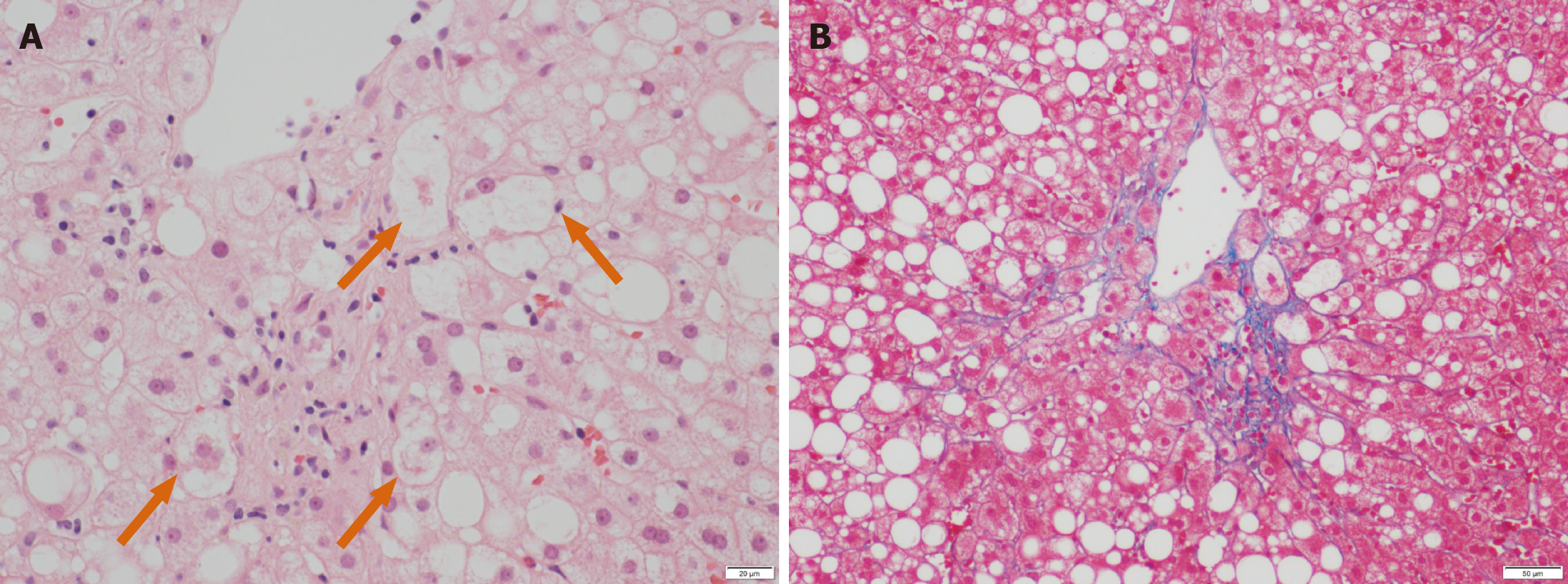
Figure 1 Pathological findings of liver biopsy.
A: Liver specimen stained by hematoxylin and eosin exhibited moderate macrovesicular steatosis mainly around the central vein with occasional focal lobular inflammation. Some ballooned hepatocytes were detected (orange arrows) According to the criteria proposed by Brunt et al, the score for steatosis, lobular inflammation, and hepatocyte ballooning were 2 (moderate), 1 (few), and 1 (few), respectively. The calculated NAFLD activity score was 4. Scale bar represents 20 μm; B: Liver specimen stained by the Azan-Mallory method showed pericentral/perisinusoidal fibrosis (score 1a). Scale bar represents 50 μm.
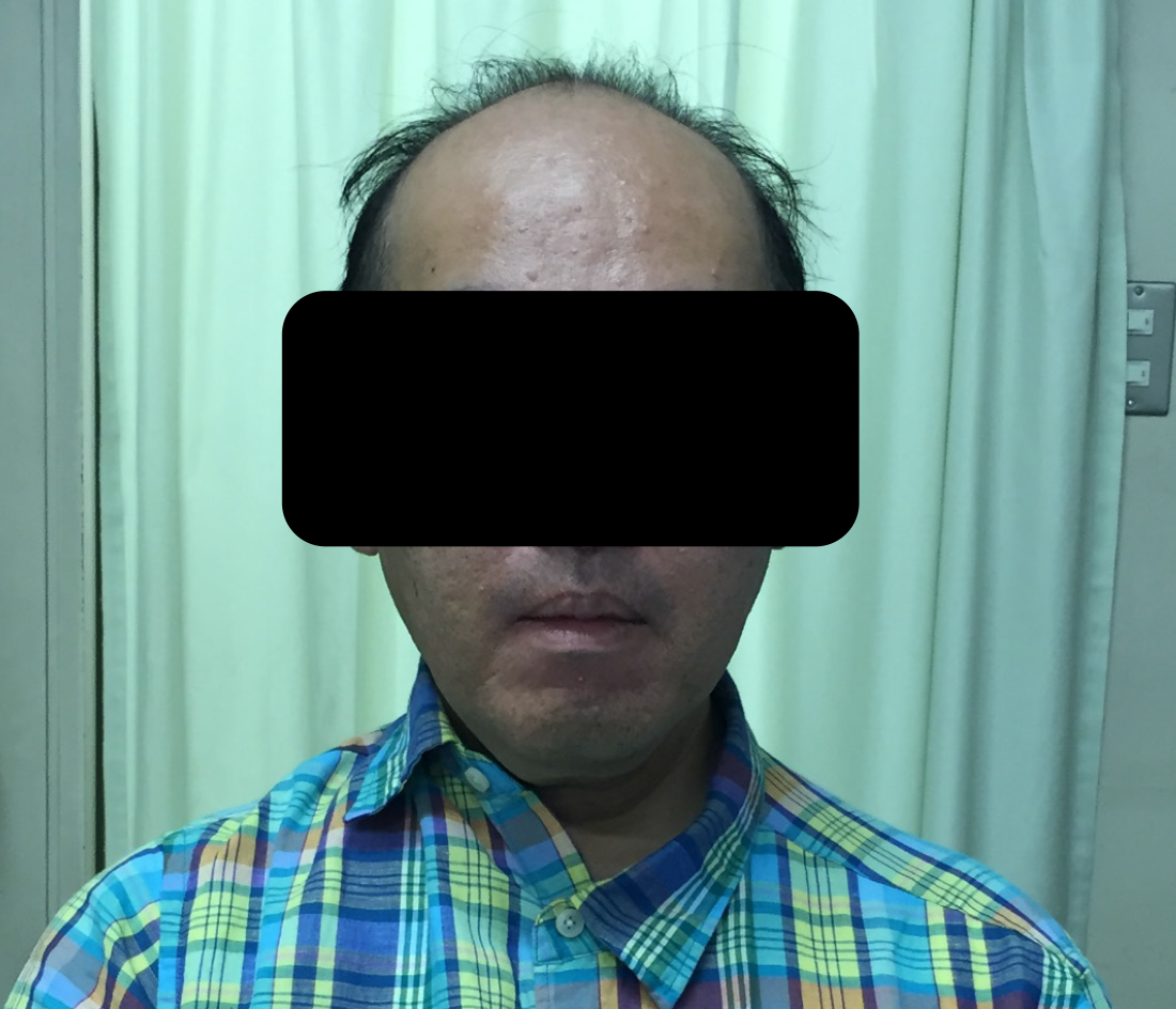
Figure 2 Photograph of the patient.
Frontal baldness and a hatchet face were noted.
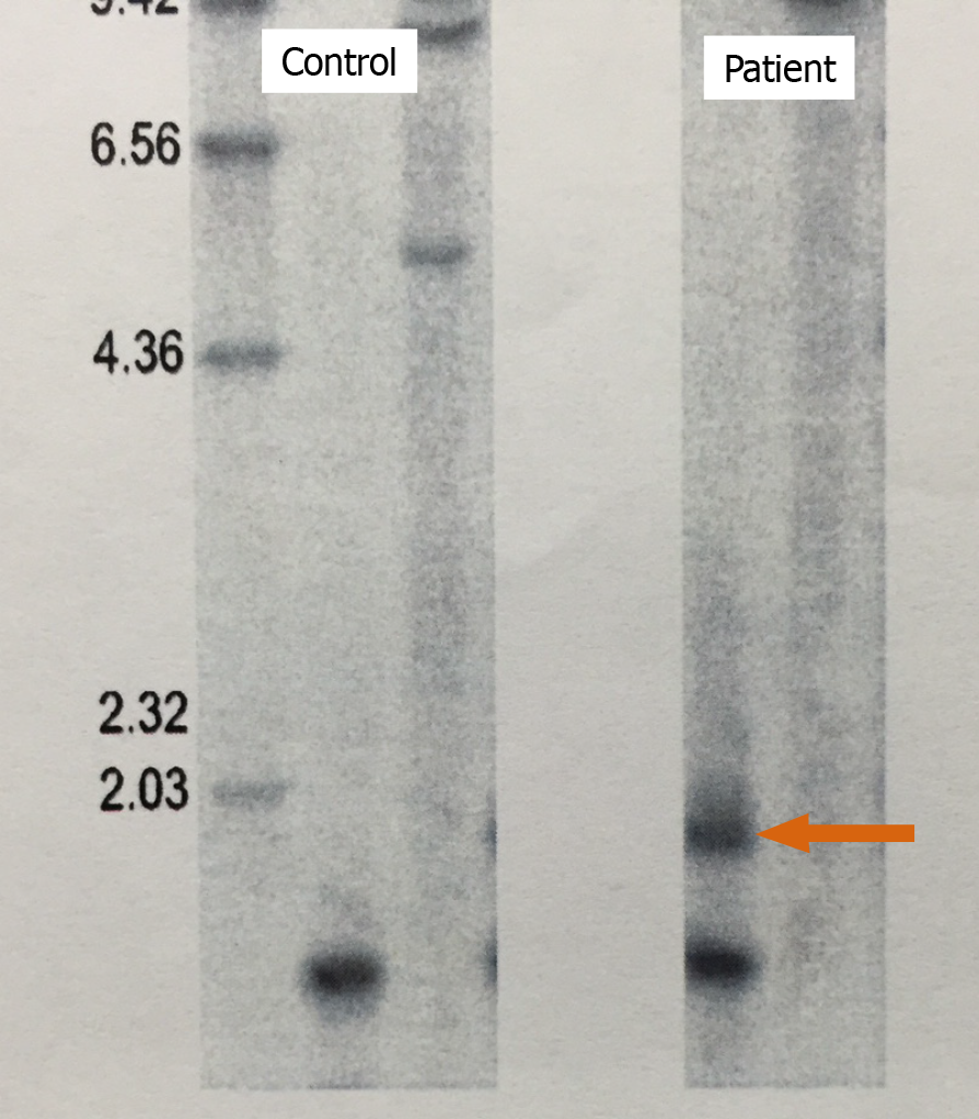
Figure 3 Southern blotting of patient DNA.
DNA samples were digested with BamHI. Compared with the control sample, an additional band caused by CTG repeats was detected (arrow), confirming the diagnosis of myotonic dystrophy.
- Citation: Tanaka N, Kimura T, Fujimori N, Ichise Y, Sano K, Horiuchi A. Non-alcoholic fatty liver disease later diagnosed as myotonic dystrophy. World J Hepatol 2020; 12(9): 685-692
- URL: https://www.wjgnet.com/1948-5182/full/v12/i9/685.htm
- DOI: https://dx.doi.org/10.4254/wjh.v12.i9.685


