Copyright
©The Author(s) 2020.
World J Hepatol. Apr 27, 2020; 12(4): 170-183
Published online Apr 27, 2020. doi: 10.4254/wjh.v12.i4.170
Published online Apr 27, 2020. doi: 10.4254/wjh.v12.i4.170
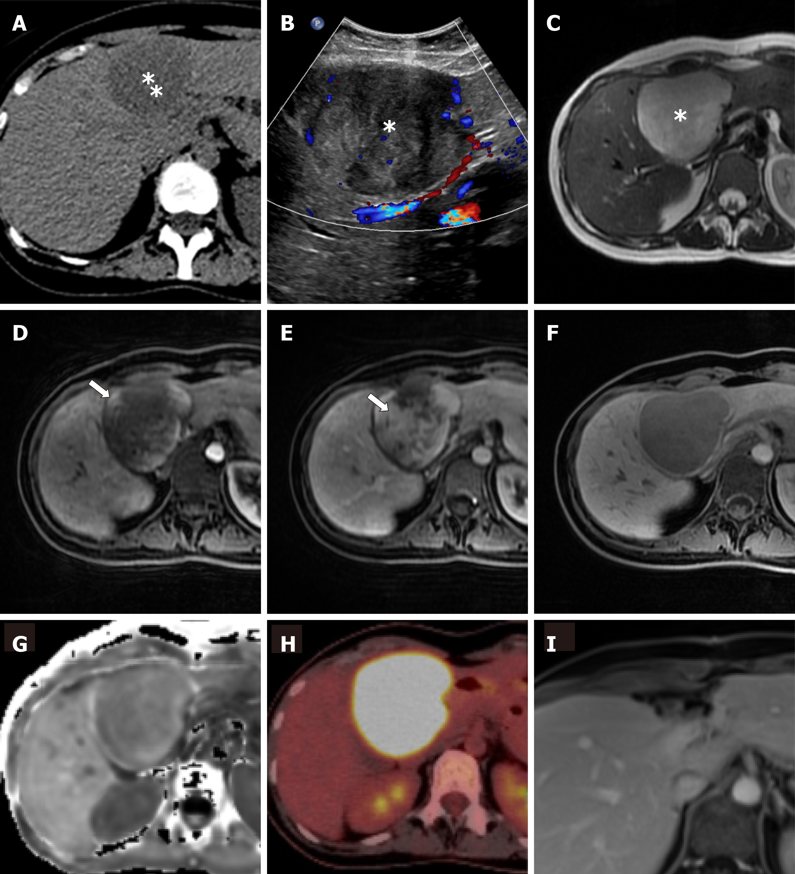
Figure 1 Imaging features within the liver lesion in segment IV.
A: The lesion was a first detected as an incidental finding in an unenhanced abdominal computed tomography to rule out kidney stones (asterisk); B: Conformed with an ultrasound examination (asterisk); C: In a following magnetic resonance imaging the lesion showed a homogeneous high signal in T2-weighted imaging (asterisk); D: After the application of intravenous hepatocyte specific contrast medium (gadoxetic acid, Primovist®/Eovist®, Bayer Healthcare Pharmaceuticals, Leverkusen, Germany) there was an early enhancement at the rim in the arterial phase (arrow); E: Followed by a strong enhancement in the venous phase (arrow); F: In the hepatobiliary phase after 20 min, the lesion appeared with a low intracellular uptake of the contrast medium compared with the adjacent liver tissue; G: In the diffusion-weighted imaging there was no clear diffusion restriction detection within the lesion (apparent diffusion coefficient); H: In an additional positron emission tomography-computed tomography examination the lesion showed an intensively increased tracer uptake; I: A follow-up magnetic resonance imaging examination after 3 mo confirmed a complete surgical resection (with multiple artifacts at the resection margin due to multiple clips) and ruled out new hepatic lesions.
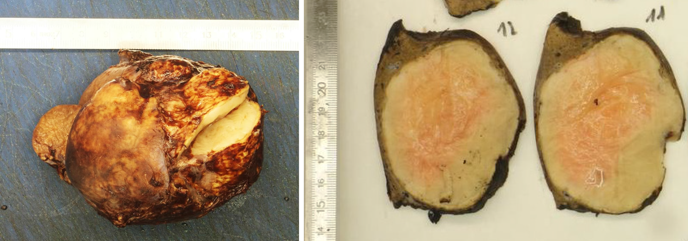
Figure 2 Postoperative macroscopic pathology of the inflammatory myofibroblastic tumors.
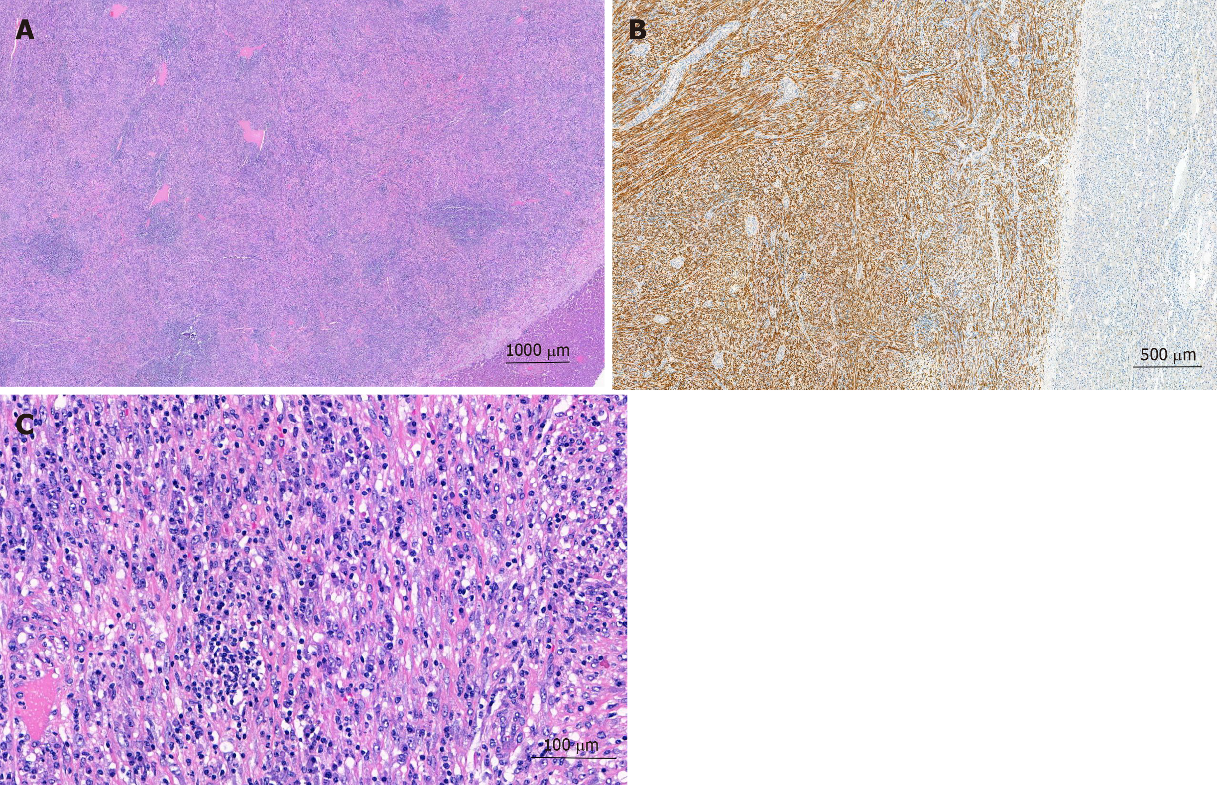
Figure 3 Postoperative microscopic pathology of the inflammatory myofibroblastic tumors.
A: Well demarcated firm vascularized tumor mass with spotty inflammatory infiltrate; B: Bland proliferation of spindle cells in broad fascicles at higher magnification. Scattered lymphocytes and plasma cell; C: Intense positivity of the spindel cells for anaplastic lymphoma kinase.
- Citation: Filips A, Maurer MH, Montani M, Beldi G, Lachenmayer A. Inflammatory myofibroblastic tumor of the liver: A case report and review of literature. World J Hepatol 2020; 12(4): 170-183
- URL: https://www.wjgnet.com/1948-5182/full/v12/i4/170.htm
- DOI: https://dx.doi.org/10.4254/wjh.v12.i4.170


