Copyright
©The Author(s) 2020.
World J Hepatol. Feb 27, 2020; 12(2): 64-71
Published online Feb 27, 2020. doi: 10.4254/wjh.v12.i2.64
Published online Feb 27, 2020. doi: 10.4254/wjh.v12.i2.64
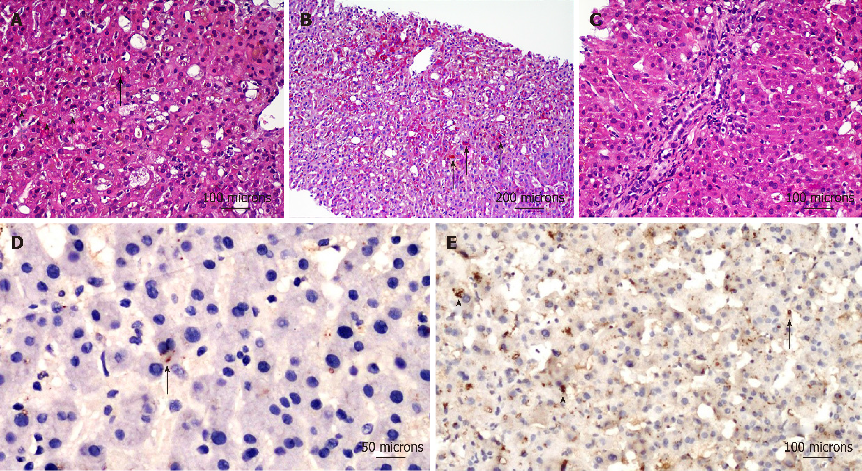
Figure 1 Histological pictures of liver biopsy and immunohistochemical analysis of ATP8B1 gene encoded protein in liver biopsy.
A: Severe cholestasis in zone 3 (centrilobular area) with evidence of bile plugs (black arrow) as well as intra-hepatocyte bile pigment [hematoxylin and eosin (HE), 200 ×]; B: Activation and hyperplasia of Kupffer’s cells, which are stained red by periodic acid schiff (PAS) reaction (black arrow) and counterstained blue by haematoxylin (PAS, 100 ×); C: Portal area showing preserved biliary duct and few inflammatory cells (HE, 200 ×); D: Canalicular immunoreactivity (diaminobenzidine chromogen and hematoxylin counterstain) for ATP8B1 gene encoded protein showing mild (black arrow) or absent staining in our benign recurrent intrahepatic cholestasis patient (400 ×); E: Normal immunohistochemical ATP8B1 gene encoded protein (black arrow) staining in a healthy control (200 ×). Scale bars with length expressed as microns are reported.
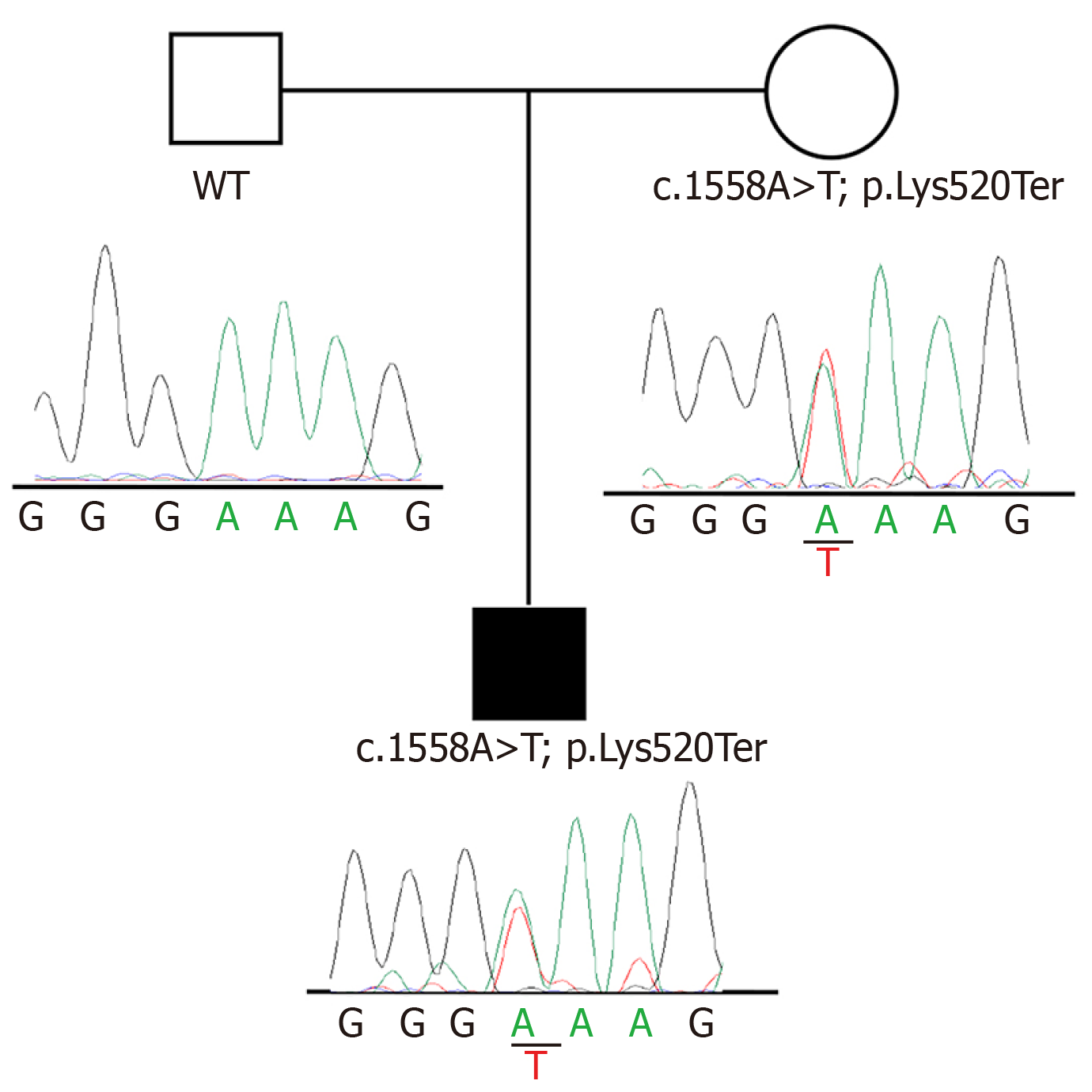
Figure 2 Pedigree and electropherograms of ATP8B1 c.
1558A>T variant identified in the family of the BRIC patient.
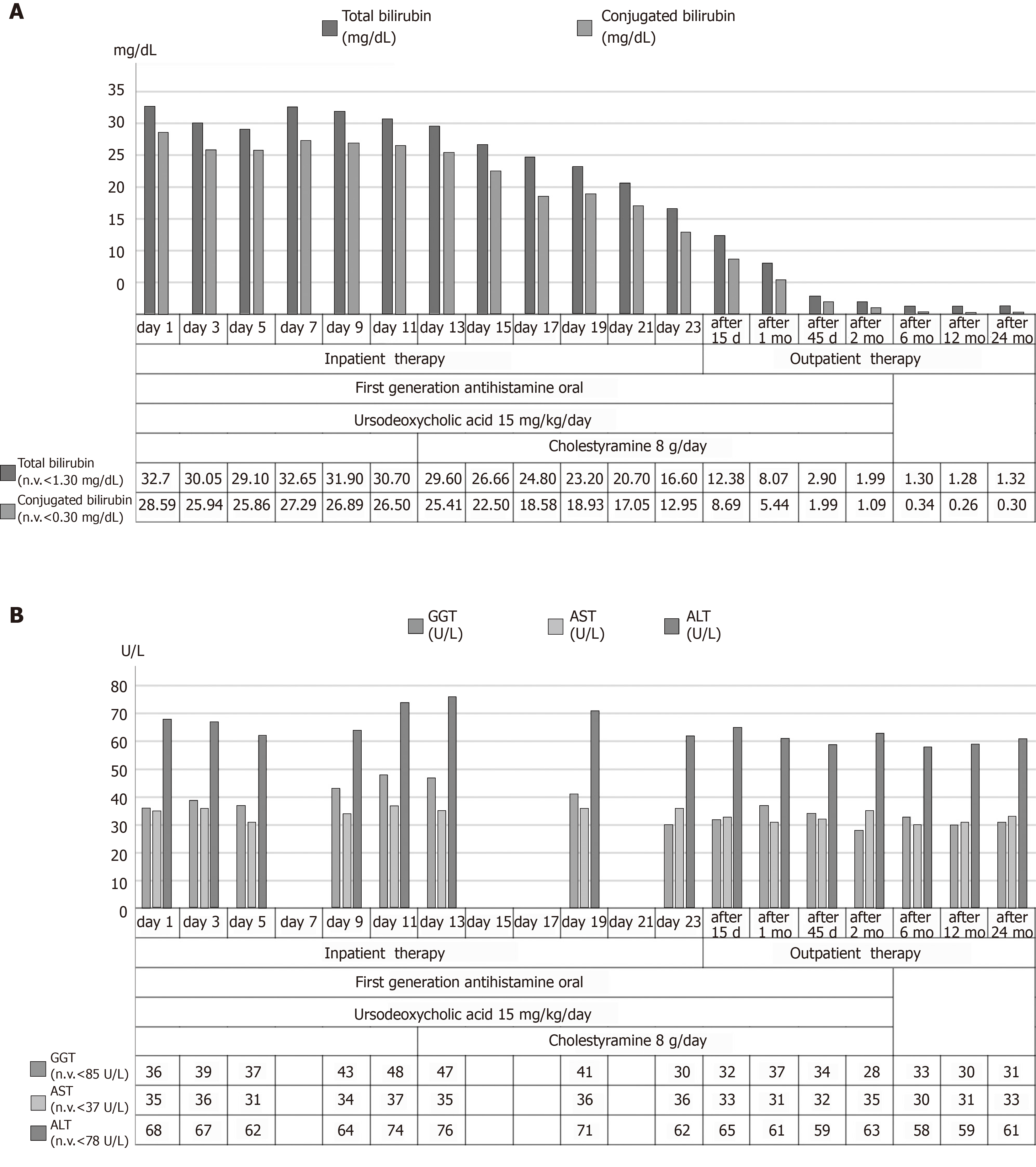
Figure 3 Course of most significant laboratory tests and trend chart with the therapeutic process.
A: Total and conjugated bilirubin values, expressed as mg/dL; B: Aspartate aminotransferase, alanine aminotransferase and gamma-glutamyltranspeptidase, expressed as U/L. n.v.: Normal values; GGT: Gamma-glutamyltranspeptidase; AST: Aspartate aminotransferase; ALT: Alanine aminotransferase.
- Citation: Piazzolla M, Castellaneta N, Novelli A, Agolini E, Cocciadiferro D, Resta L, Duda L, Barone M, Ierardi E, Di Leo A. Nonsense variant of ATP8B1 gene in heterozygosis and benign recurrent intrahepatic cholestasis: A case report and review of literature. World J Hepatol 2020; 12(2): 64-71
- URL: https://www.wjgnet.com/1948-5182/full/v12/i2/64.htm
- DOI: https://dx.doi.org/10.4254/wjh.v12.i2.64


