Copyright
©The Author(s) 2020.
World J Hepatol. Oct 27, 2020; 12(10): 863-869
Published online Oct 27, 2020. doi: 10.4254/wjh.v12.i10.863
Published online Oct 27, 2020. doi: 10.4254/wjh.v12.i10.863
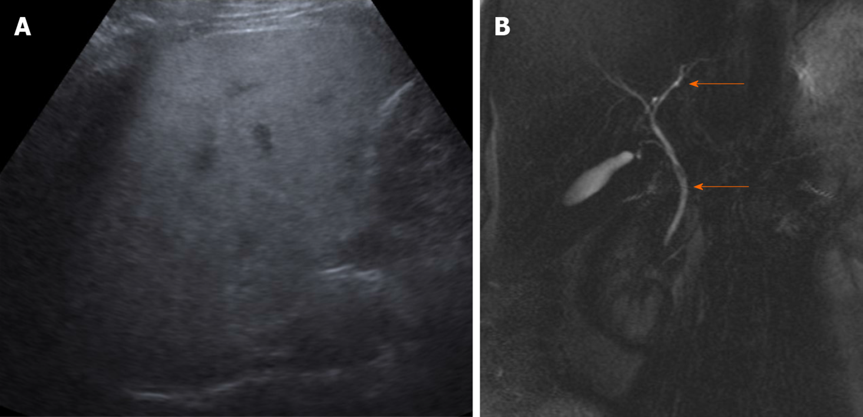
Figure 1 The abdominal ultrasound.
A: Diffuse fatty infiltration of liver; B: Normal common bile duct and intrahepatic biliary ducts (orange arrows) in magnetic resonance cholangiopancreatography 3D image.
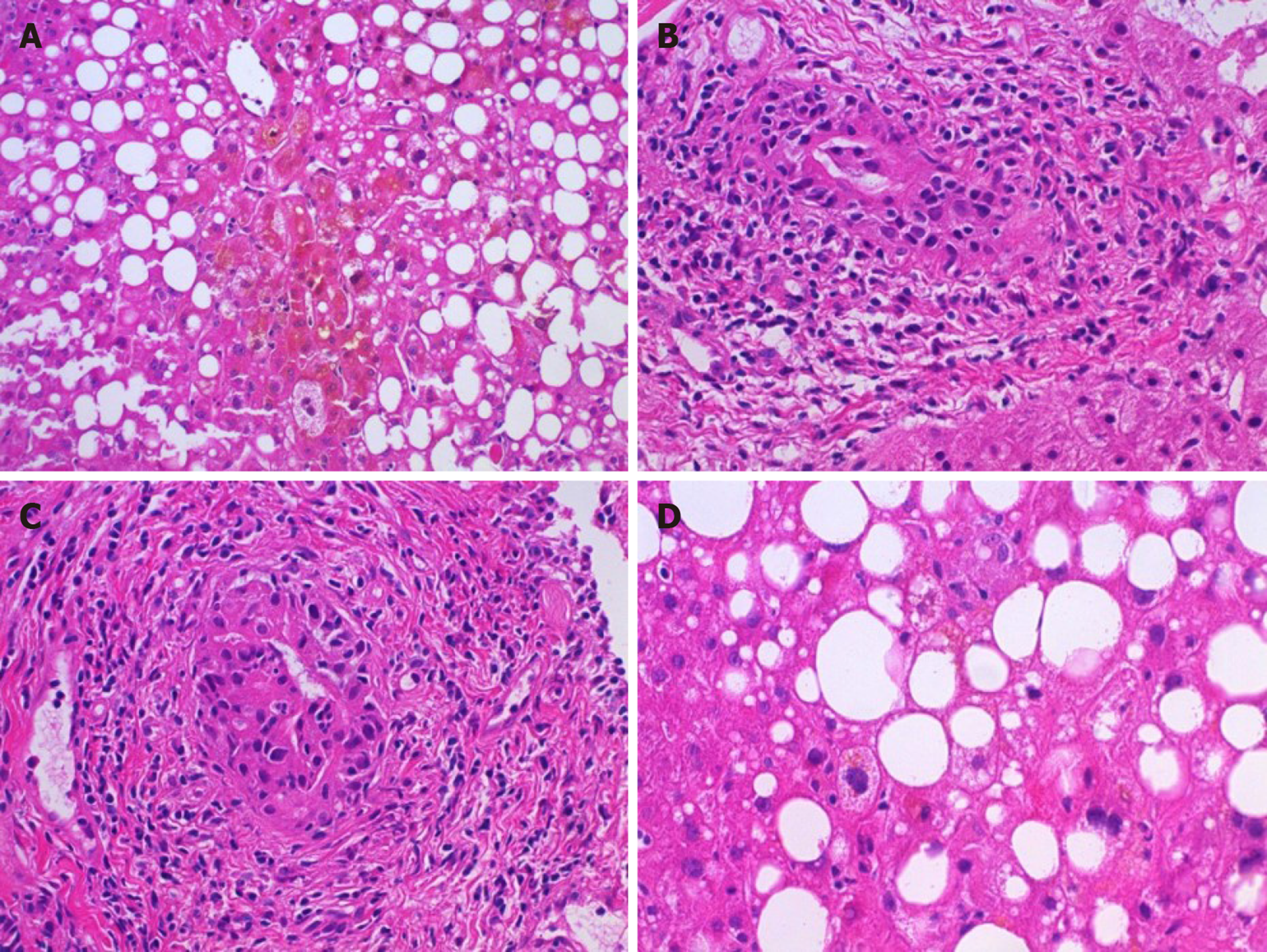
Figure 2 Histopathological findings.
A: Centrilobular areas showed well defined cholestasis; B and C: Portal tracts showed moderate chronic inflammation and brisk lymphocytic-predominant bile duct injury; D: Background liver showed steatohepatitis which was felt to most likely be due to underlying obesity-related non-alcoholic fatty liver disease.
- Citation: Gandhi D, Ahuja K, Quade A, Batts KP, Patel L. Kratom induced severe cholestatic liver injury histologically mimicking primary biliary cholangitis: A case report. World J Hepatol 2020; 12(10): 863-869
- URL: https://www.wjgnet.com/1948-5182/full/v12/i10/863.htm
- DOI: https://dx.doi.org/10.4254/wjh.v12.i10.863


