Copyright
©The Author(s) 2019.
World J Hepatol. Apr 27, 2019; 11(4): 402-408
Published online Apr 27, 2019. doi: 10.4254/wjh.v11.i4.402
Published online Apr 27, 2019. doi: 10.4254/wjh.v11.i4.402
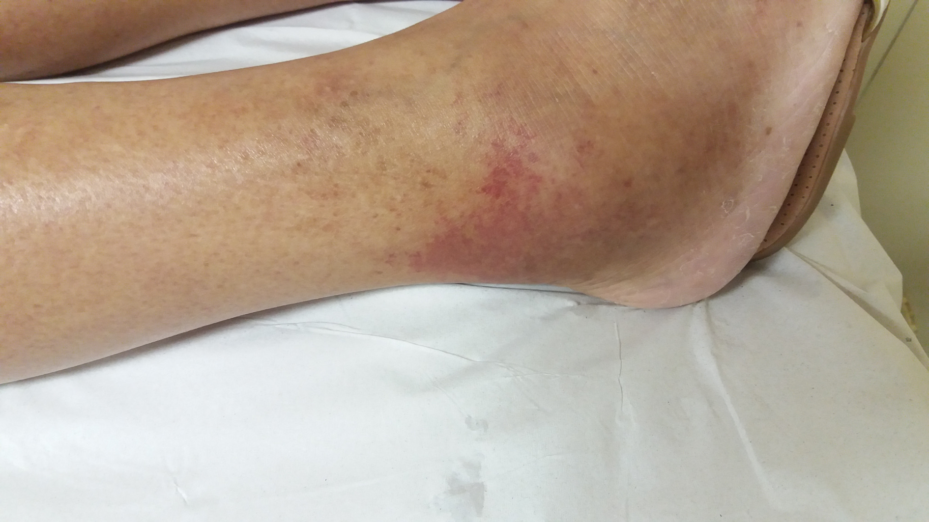
Figure 1 Aspect of the lesion on the patient´s right foot.
The image was taken on the day she presented to the emergency room.
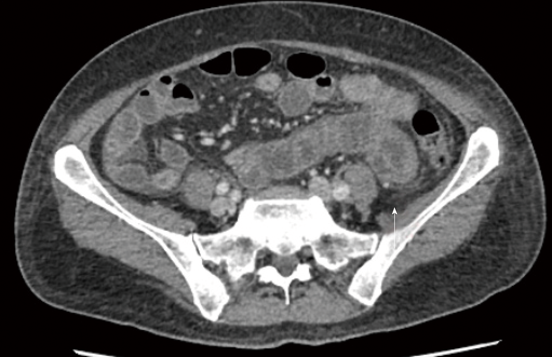
Figure 2 Computerized tomography scan showing a small volume of ascites and diffuse thickening of bowel walls (white arrow).
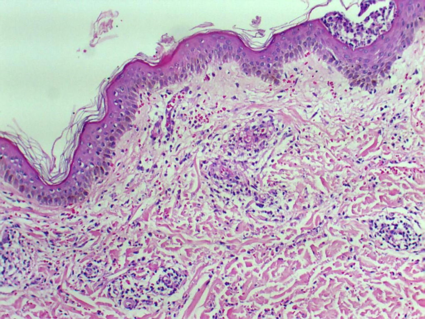
Figure 3 Focal vascular damage with mild perivascular neutrophilic infiltrate and fragmentation of the neutrophils resulting in nuclear dust (leukocytoclasis), suggestive of Urticarial vasculitis (HE 100 x).
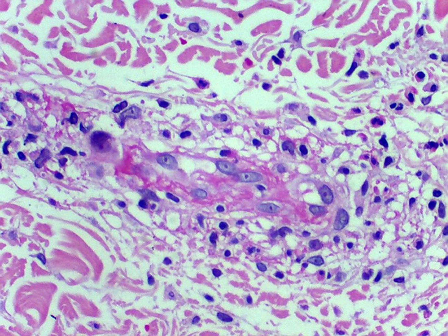
Figure 4 Perivascular neutrophilic infiltrate (HE 40 x).
- Citation: Ferreira GSA, Watanabe ALC, Trevizoli NC, Jorge FMF, Diaz LGG, Araujo MCCL, Araujo GC, Machado AC. Leukocytoclastic vasculitis caused by hepatitis C virus in a liver transplant recipient: A case report. World J Hepatol 2019; 11(4): 402-408
- URL: https://www.wjgnet.com/1948-5182/full/v11/i4/402.htm
- DOI: https://dx.doi.org/10.4254/wjh.v11.i4.402


