Copyright
©The Author(s) 2019.
World J Hepatol. Mar 27, 2019; 11(3): 294-304
Published online Mar 27, 2019. doi: 10.4254/wjh.v11.i3.294
Published online Mar 27, 2019. doi: 10.4254/wjh.v11.i3.294
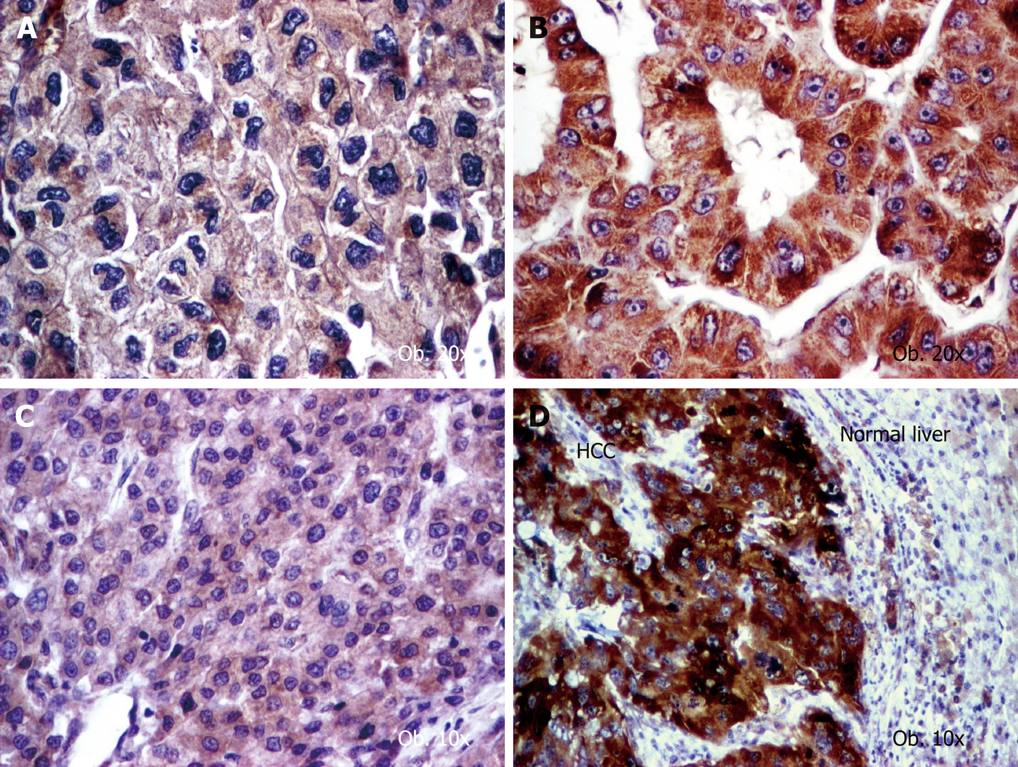
Figure 1 Angiogenic phenotype of hepatocellular carcinoma cells.
A: Vascular endothelial growth factor (VEGF) A shows a low intensity (score 2+) in aggressive cases with vascular invasion; B: VEGF-A is well expressed (score 3+) in well differentiated carcinomas; C: Cyclooxygenase-2 (COX-2) presents low intensity (score 1+) in well-differentiated carcinomas; D: COX-2 becomes upregulated (score 3+) in dedifferentiated tumors.
Figure 2 Particular features of neoangiogenesis of hepatocellular carcinoma, revealed by CD31 stain.
A: Endothelization of the cirrhotic synusoids; B: High endothelial area and immature vessels in the peri-cirrhotic tumor tissue; C: Rare mature vessels can be intratumorally seen, in cases developed in patients without cirrhosis. 20 x.
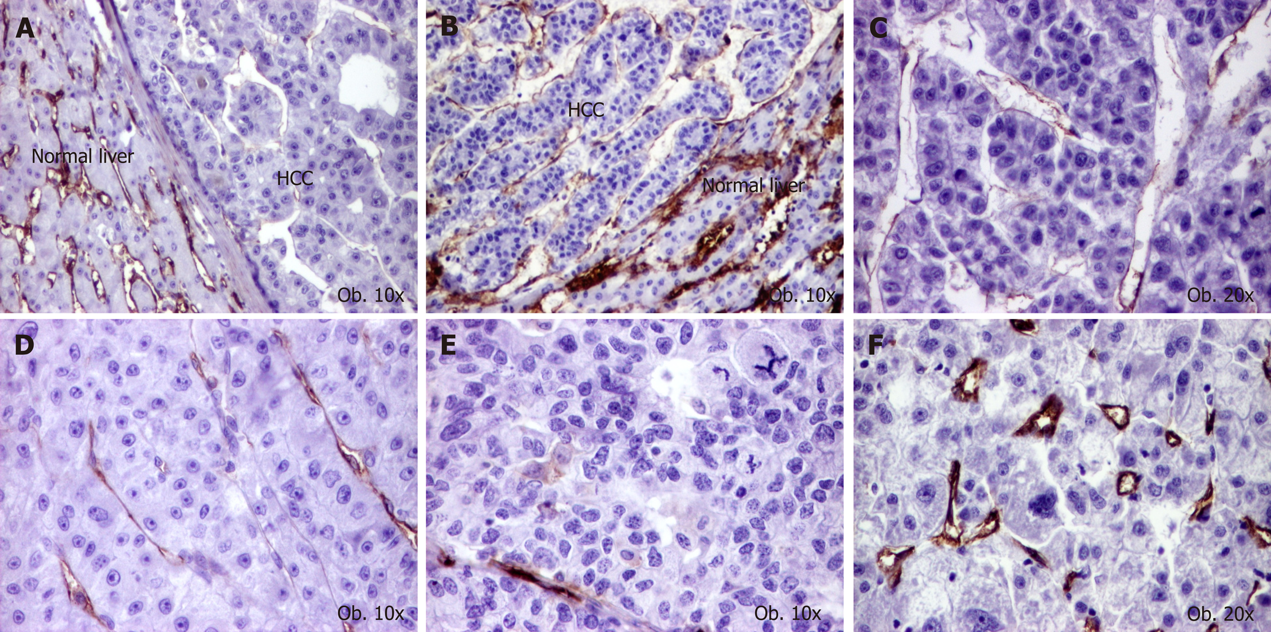
Figure 3 Particular features of neoangiogenesis of hepatocellular carcinoma, revealed by CD105 stain.
A and B: High microvessels density of normal parenchyma, compared with tumor tissue; C: Mature activated neoformed vessels, in a G1 carcinoma; D: Mature vessels, in a G2 carcinoma; E: Low endothelial area, in a G3 carcinoma; F: Small neoformed vessels, in a case with high endothelial area.
- Citation: Fodor D, Jung I, Turdean S, Satala C, Gurzu S. Angiogenesis of hepatocellular carcinoma: An immunohistochemistry study. World J Hepatol 2019; 11(3): 294-304
- URL: https://www.wjgnet.com/1948-5182/full/v11/i3/294.htm
- DOI: https://dx.doi.org/10.4254/wjh.v11.i3.294


