Copyright
©The Author(s) 2019.
World J Hepatol. Jan 27, 2019; 11(1): 127-132
Published online Jan 27, 2019. doi: 10.4254/wjh.v11.i1.127
Published online Jan 27, 2019. doi: 10.4254/wjh.v11.i1.127
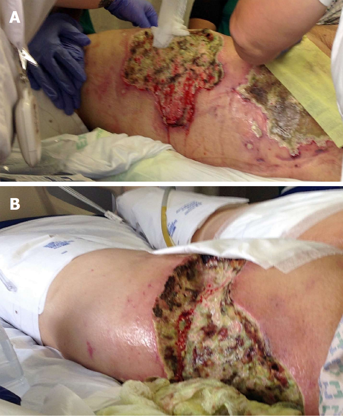
Figure 1 Calciphylaxis wounds in the thigh.
A: Before debridement; B: After debridement.
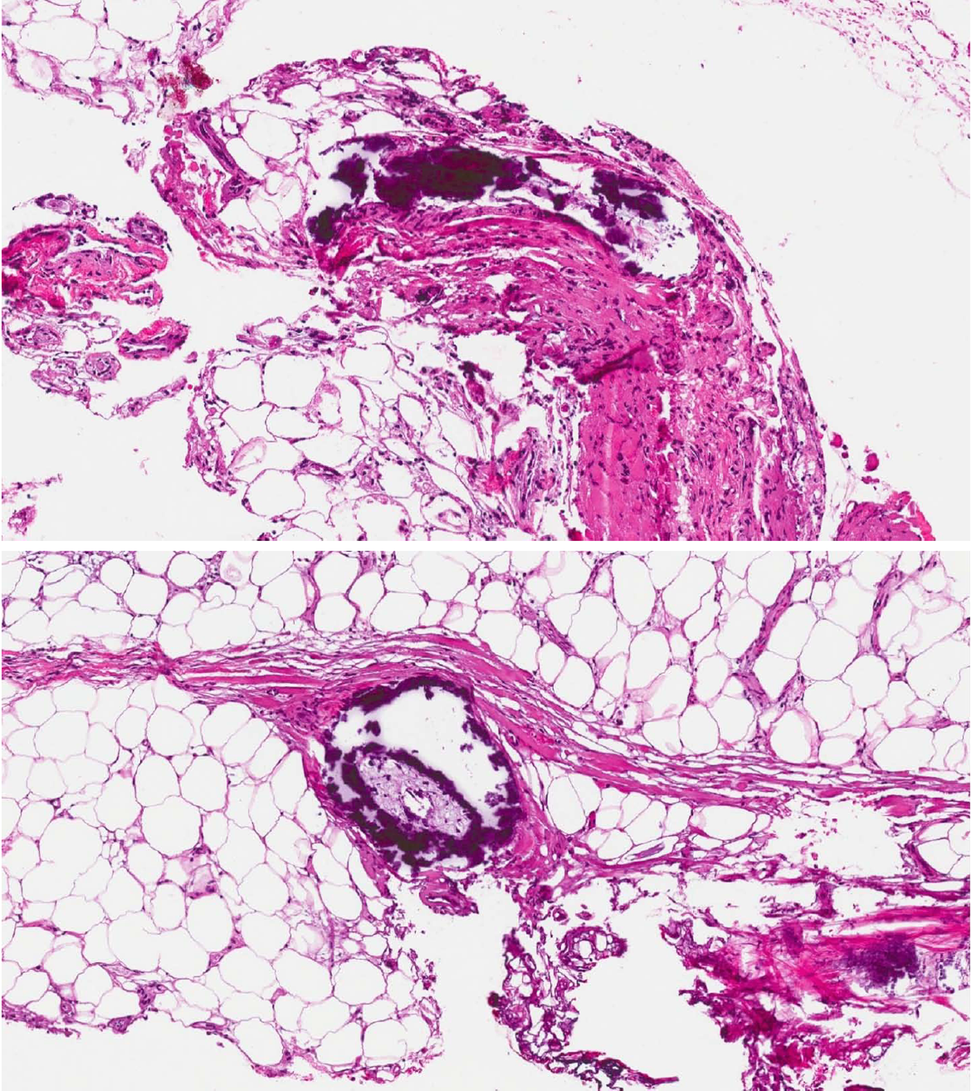
Figure 2 Dermal vascular occlusion and calcium deposition within the walls of large veins and the surrounding adipose tissue.
- Citation: Sammour YM, Saleh HM, Gad MM, Healey B, Piliang M. Non-uremic calciphylaxis associated with alcoholic hepatitis: A case report. World J Hepatol 2019; 11(1): 127-132
- URL: https://www.wjgnet.com/1948-5182/full/v11/i1/127.htm
- DOI: https://dx.doi.org/10.4254/wjh.v11.i1.127


