Copyright
©The Author(s) 2018.
World J Hepatol. Sep 27, 2018; 10(9): 629-636
Published online Sep 27, 2018. doi: 10.4254/wjh.v10.i9.629
Published online Sep 27, 2018. doi: 10.4254/wjh.v10.i9.629
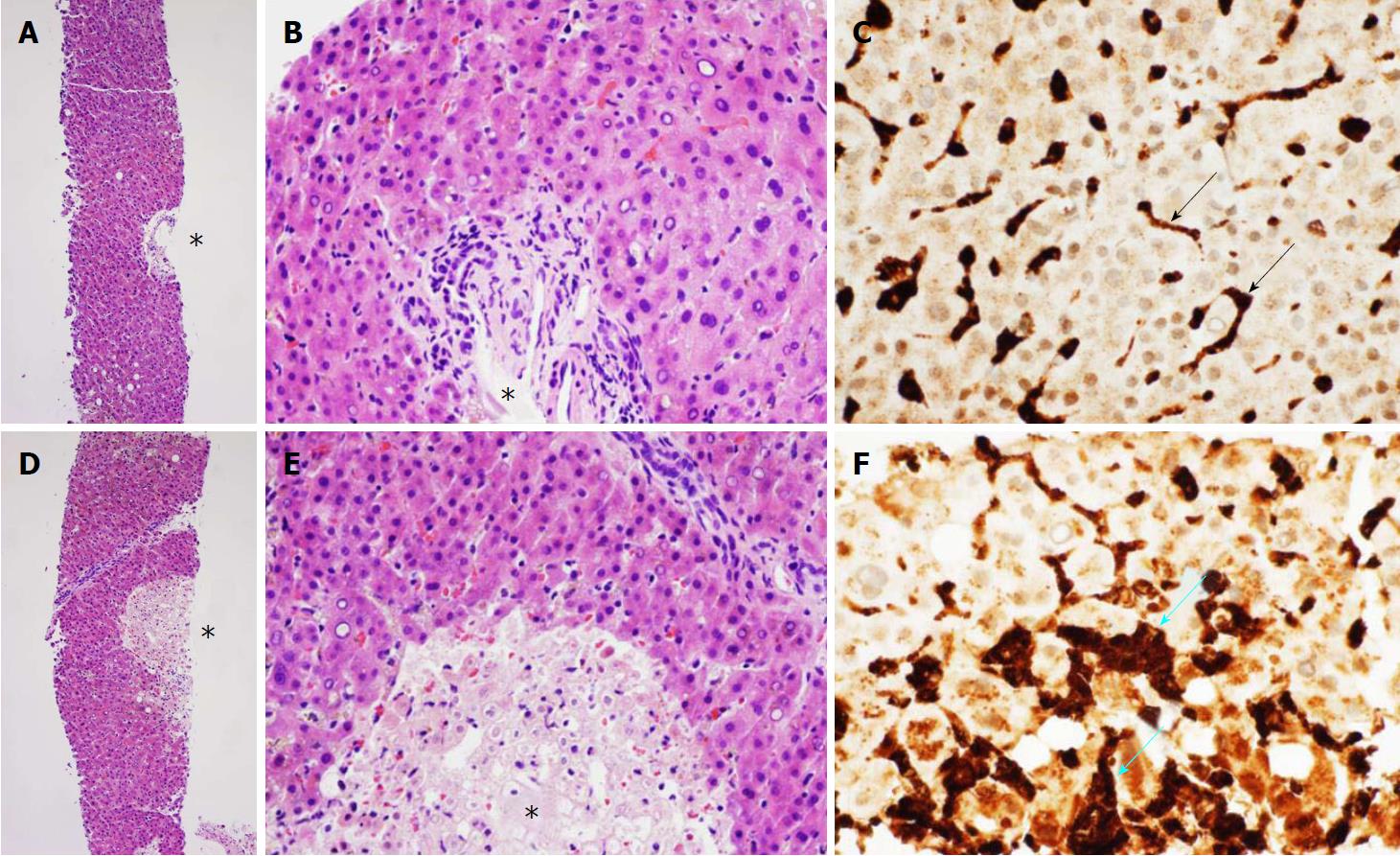
Figure 1 Liver biopsy.
A, B: Low-power (A) and high-power (B) photomicrographs of a representative area of hematoxylin and eosin-stained section of the transjugular liver needle biopsy shows a normal portal triad (asterisk) and viable and relatively normal appearing hepatocytes with characteristically deeply eosinophilic cytoplasm and without evidence of hepatocyte necrosis, lymphocytic infiltrates, macrophages or hemophagocytic macrophages/histiocytes; C: Medium power photomicrograph with immunohistochemistry for CD-68, a molecular marker for Kupffer cells and tissue macrophages, shows CD-68 positive (brown staining) cells with elongated and slender morphology characteristic of Kupffer cells (arrows) within hepatic sinusoids; D, E: Low-power (D) and high-power (E) photomicrographs of hematoxylin and eosin-stained section of liver biopsy exhibiting focal centrilobular necrosis (asterisk), seen as a pale, circumscribed area containing pyknotic nuclei in liver biopsy specimen. Despite the liver failure, the areas of necrosis were sparsely distributed, and the overwhelming majority of the liver biopsy specimen contained viable and relatively well-preserved hepatocytes except for the cholestasis, demonstrated by a faint yellowish-brown tint in hepatocytes. No lymphocytic infiltrates or hemophagocytic macrophages/histiocytes are detected in this H and E stain; F: Medium-power photomicrograph with immunohistochemistry for CD-68, a molecular marker for Kupffer cells and macrophages, shows CD-68-positive (brown staining) plump cells with irregular contours characteristic of macrophages (blue arrows). The macrophages have been recruited to phagocytose the dying hepatocytes in the focally ischemic centrilobular area.
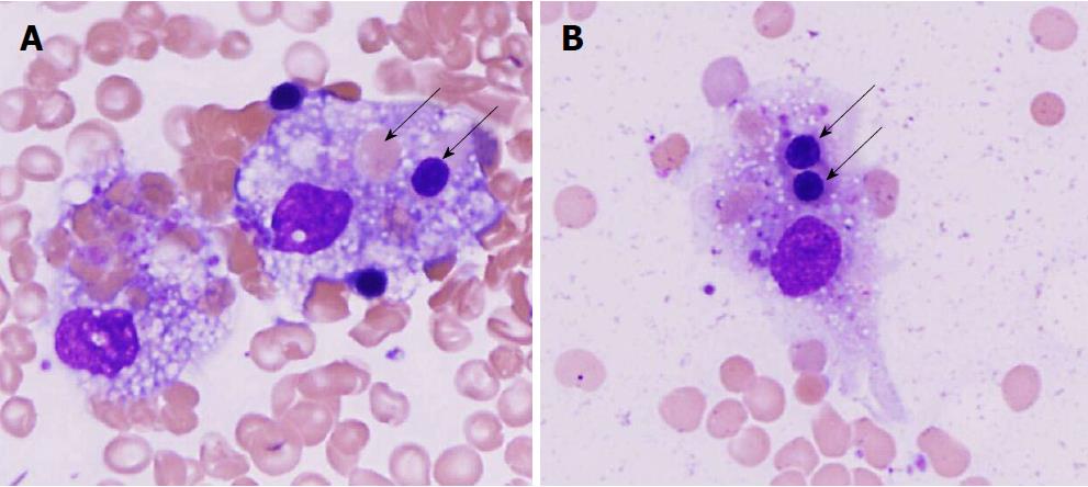
Figure 2 Hemophagocytosis in bone marrow.
A: High-power photomicrograph of Diff Quick stain of bone marrow smear showing two large macrophages with foamy cytoplasm within a background of mature erythrocytes. The macrophage on the right has phagocytosed a mature erythrocyte (pink cell with no nucleus, higher arrow) and a nucleated erythrocyte precursor (pink cell with deeply purple nucleus, lower arrow; B: high-power photomicrograph of another large macrophage with foamy cytoplasm containing two phagocytosed nucleated erythrocyte precursor cells (purple nucleus, two arrows).
- Citation: Cappell MS, Hader I, Amin M. Acute liver failure secondary to severe systemic disease from fatal hemophagocytic lymphohistiocytosis: Case report and systematic literature review. World J Hepatol 2018; 10(9): 629-636
- URL: https://www.wjgnet.com/1948-5182/full/v10/i9/629.htm
- DOI: https://dx.doi.org/10.4254/wjh.v10.i9.629


