Copyright
©The Author(s) 2018.
World J Hepatol. Aug 27, 2018; 10(8): 530-542
Published online Aug 27, 2018. doi: 10.4254/wjh.v10.i8.530
Published online Aug 27, 2018. doi: 10.4254/wjh.v10.i8.530
Figure 1 Pathological changes of liver simple steatosis and cirrhosis.
A: 45-year-old man with simple steatosis. The liver biopsy shows marked macrovesicular steatosis without inflammation or fibrosis (H and E x4); B: 48-year-old man with cirrhosis due to non-alcoholic fatty liver disease. In addition to marked macrovesicular steatosis, there is loss of normal hepatic architecture and replacement by regenerative nodules surrounded by bands of fibrous tissue, a characteristic feature of cirrhosis (H and E x4).
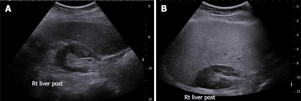
Figure 2 Grey-scale ultrasound in non-alcoholic fatty liver disease.
A: 47-year-old female with increased echogenicity of the liver relative to the right kidney, a classic sonographic finding of hepatic steatosis. The patient had elevated serum liver enzymes and underwent a liver biopsy for NASH evaluation; B: 51-year-old female who underwent liver biopsy as part of clinical follow-up. On evaluation of liver pathology, there was no steatosis. Ultrasound image shows normal echogenicity of the liver parenchyma, which is only slightly hyperechoic relative to the renal parenchyma.
Figure 3 Share wave elastography in non-alcoholic fatty liver disease.
A: 54-year old female who underwent liver biopsy for NASH evaluation. The biopsy demonstrated steatosis only with no inflammation or fibrosis. SWE median value of 7.05 kPa; B: SWE in a 52-year old female that underwent liver biopsy for the evaluation of NASH. A median SWE value of 11.5 kPa was significantly higher than normal liver with steatosis only. On biopsy, the subject had fibrosis stage 0 according to the METAVIR system and fibrosis stage 1a as per the NAS CRN criteria. The non-alcoholic fatty liver disease activity score in this patient was 6, consistent with NASH. NASH: Non-alcoholic steatohepatitis; SWE: Share wave elastography.
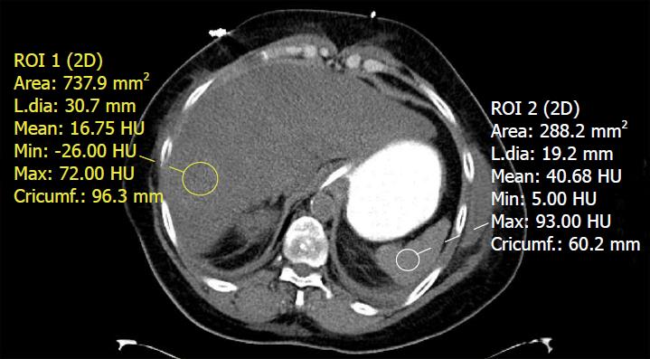
Figure 4 Unenhanced computed tomography of the abdomen in a patient with fatty liver disease.
Regions of interest placed within the liver and spleen demonstrate a hepatic attenuation of 16.75 Hounsfield units (less than 40) and a splenic attenuation of 40.68 Hounsfield units. This meets the definition of fatty liver on CT by absolute value, liver/spleen attenuation difference, and liver/spleen attenuation ratio criteria.
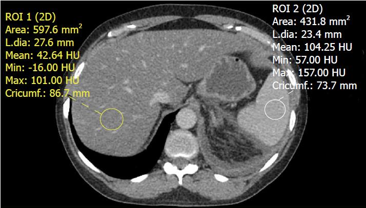
Figure 5 Contrast-enhanced computed tomography of the abdomen demonstrating a fatty liver.
The liver has an attenuation value of 42.64 Hounsfield units while the spleen has an attenuation value of 104.25 Hounsfield units. An attenuation difference of 62 HU is highly suggestive of fatty liver disease.
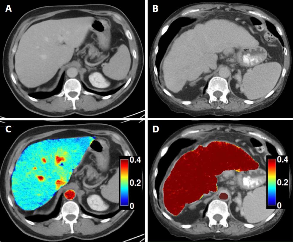
Figure 6 Dual-energy CT images for the assessment of liver fibrosis.
A: Delayed phase axial CT images from a patient with mild fibrosis; B: Severe fibrosis; C-D: DECT color overlay contrast agent maps. Iodine concentration within the liver parenchyma in reference to that in the aorta [NIC (normalized iodine concentration) in mg/mL = I Liver / I Aorta] on 5 min delayed acquisitions can be seen on the images. Since contrast media is retained within fibrotic tissues, the NIC on delayed-phase images increases with the severity of liver fibrosis; D: Patients with severe cirrhosis have higher parenchymal contrast media retention on delayed images in relationship to the aorta, as compared to the mild retention in patients with lesser grades of liver fibrosis (C).
Figure 7 Hepatic steatosis on magnetic resonance imaging.
A, B: In phase (A) and out of phase (B) gradient echo T1-weighted images of the abdomen demonstrate signal dropout on the out of phase image due to the presence of microscopic (intracellular) fat deposition in the liver; C: A fat only image via the Dixon method demonstrates diffuse fat accumulation as evidenced by increased T1 signal within the liver.
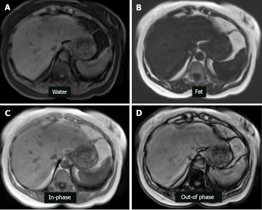
Figure 8 Magnetic resonance imaging dixon technique in the patient with hepatic steatosis.
A: Water only sequence; B: Fat only sequence; C: In-phase sequence; D: Out-of-phase sequence.
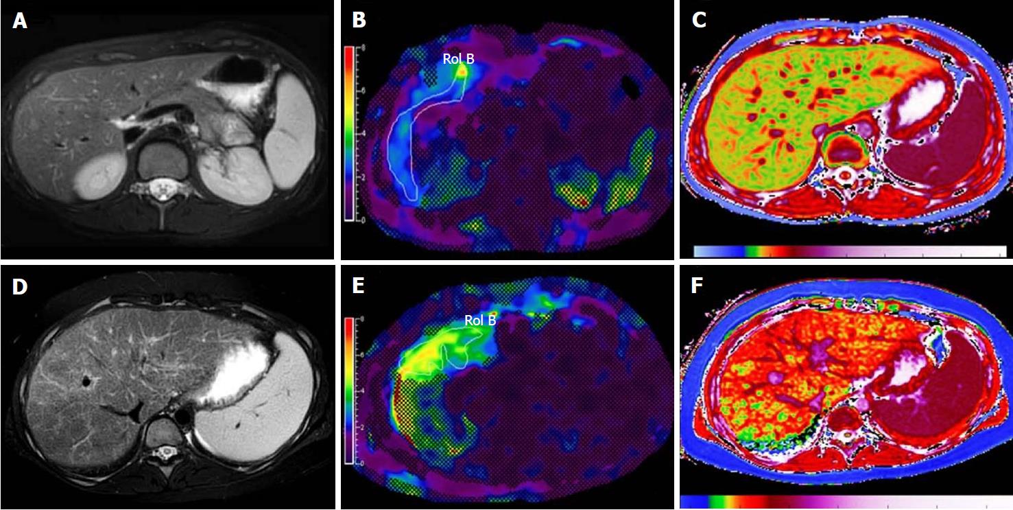
Figure 9 Examples of multiparametric magnetic resonance imaging measurements in pediatric patients[87].
A-C: MRI images in a healthy child without evidence of liver disease. A: Conventional T2-weighted fast spin echo (A); B: MRE with normal hepatic shear stiffness of 2.22 kPa (B), and corrected T1 time of 879 ms (C); D, F: Corresponding images in a child with biopsy-confirmed primary sclerosing cholangitis. D: The T2-weighted fast spin echo image shows heterogeneously increased liver signal due to fibrosis that can be quantified using texture mapping; E: MRE image shows an increased hepatic shear stiffness of 3.96 kPa; F: The corrected T1 time was increased to 1048 ms, likely due to a combination of fibrosis and inflammation.
- Citation: Li Q, Dhyani M, Grajo JR, Sirlin C, Samir AE. Current status of imaging in nonalcoholic fatty liver disease. World J Hepatol 2018; 10(8): 530-542
- URL: https://www.wjgnet.com/1948-5182/full/v10/i8/530.htm
- DOI: https://dx.doi.org/10.4254/wjh.v10.i8.530


