Copyright
©The Author(s) 2018.
World J Hepatol. May 27, 2018; 10(5): 417-424
Published online May 27, 2018. doi: 10.4254/wjh.v10.i5.417
Published online May 27, 2018. doi: 10.4254/wjh.v10.i5.417
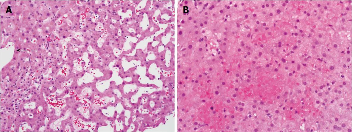
Figure 1 Sinusoidal dilatation and hepatocellular plate atrophy in zone 3 (arrow marks hepatic vein branch) (20 ×) (A); Zone 3 congestion and red blood cell extravasation (20 ×) (B).
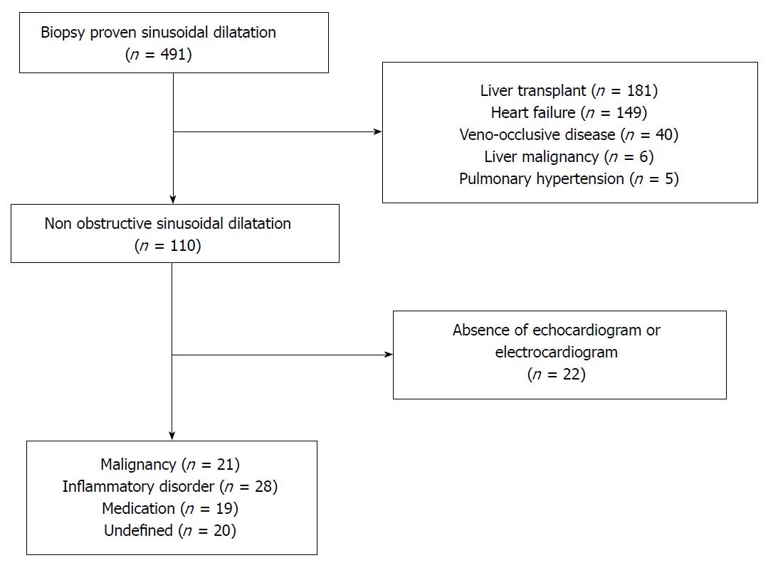
Figure 2 Flow chart of the screening process.
- Citation: Sunjaya DB, Ramos GP, Braga Neto MB, Lennon R, Mounajjed T, Shah V, Kamath PS, Simonetto DA. Isolated hepatic non-obstructive sinusoidal dilatation, 20-year single center experience. World J Hepatol 2018; 10(5): 417-424
- URL: https://www.wjgnet.com/1948-5182/full/v10/i5/417.htm
- DOI: https://dx.doi.org/10.4254/wjh.v10.i5.417


