Copyright
©2009 Baishideng.
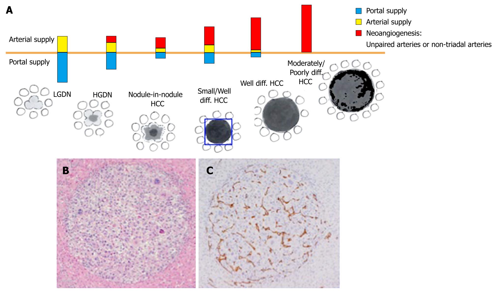
Figure 1 The diagram shows the changes of intranodular blood supply that characterises HCC (A); The sampled small/well differentiated HCC shows in HE (B) compact carcinomatous tissue well circumscribed from dysplastic tissue; CD34 immunostain of the same nodule (C) demonstrates arteries not confined to portal tracts in HCC.
diff.: Differentiated.
- Citation: Andreana L, Isgrò G, Pleguezuelo M, Germani G, Burroughs AK. Surveillance and diagnosis of hepatocellular carcinoma in patients with cirrhosis. World J Hepatol 2009; 1(1): 48-61
- URL: https://www.wjgnet.com/1948-5182/full/v1/i1/48.htm
- DOI: https://dx.doi.org/10.4254/wjh.v1.i1.48


