Copyright
©The Author(s) 2015.
World J Stem Cells. May 26, 2015; 7(4): 728-744
Published online May 26, 2015. doi: 10.4252/wjsc.v7.i4.728
Published online May 26, 2015. doi: 10.4252/wjsc.v7.i4.728
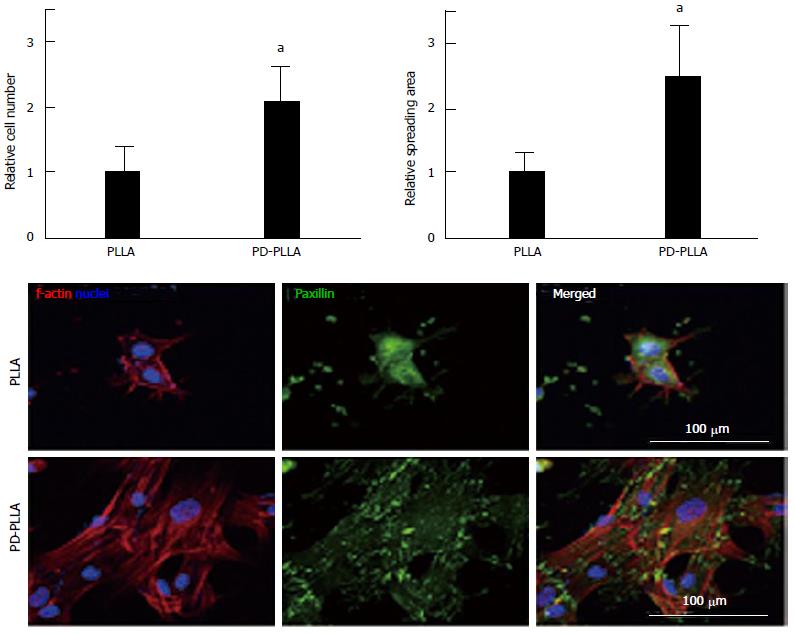
Figure 1 Quantitative analysis of initial cell adhesion from human mesenchymal stem cells cultured on the fibers.
Relative adherent cell numbers and spreading area of human mesenchymal stem cells (hMSCs) cultured on PLLA and PD-PLLA fibers were analyzed after 12 h of culture. aP < 0.05, PD-PLLA vs PLLA group. Adherent morphology of hMSCs on PLLA and PD-PLLA fibers was observed by confocal microscopy. Scale bars represent 100 μm. Reproduced with permission from Rim et al[64]. PLLA: Poly(l-lactide); PD-PLLA: Poly(l-lactide) (PLLA) fibers coated with polydopamine.
- Citation: Ghasemi-Mobarakeh L, Prabhakaran MP, Tian L, Shamirzaei-Jeshvaghani E, Dehghani L, Ramakrishna S. Structural properties of scaffolds: Crucial parameters towards stem cells differentiation. World J Stem Cells 2015; 7(4): 728-744
- URL: https://www.wjgnet.com/1948-0210/full/v7/i4/728.htm
- DOI: https://dx.doi.org/10.4252/wjsc.v7.i4.728


