Copyright
©The Author(s) 2016.
World J Stem Cells. Feb 26, 2016; 8(2): 22-31
Published online Feb 26, 2016. doi: 10.4252/wjsc.v8.i2.22
Published online Feb 26, 2016. doi: 10.4252/wjsc.v8.i2.22
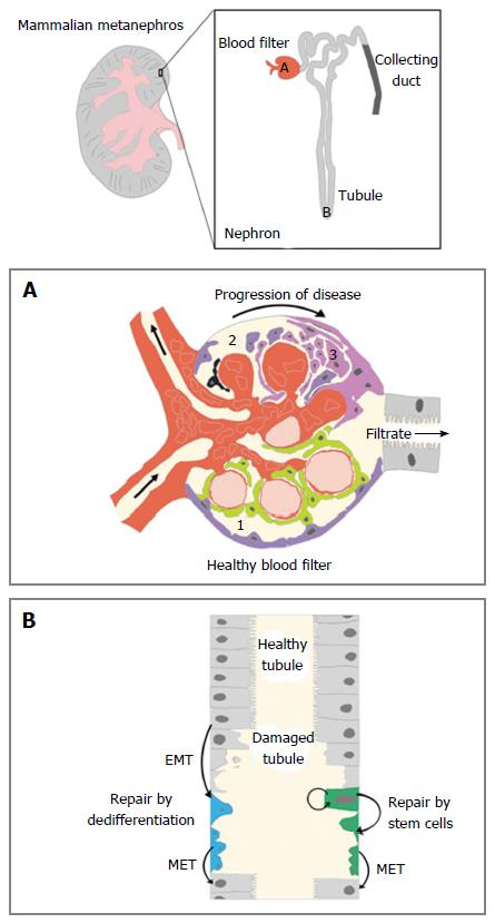
Figure 1 Kidney form, function, and regeneration.
The mammalian metanephros is composed of upwards of hundreds of thousands of small, functional subunits known as nephrons. There are three main components of the nephron: The blood filter (red), the tubule (light grey) and the collecting duct (dark grey). The unique cell types in these nephron regions have variable roles in function, injury response, and regeneration. A: (1) the healthy blood filter contains intact podocytes (green) attached to capillaries to enhance the process of blood filtration. Parietal epithelial cells or PECs (purple) that line the Bowman capsule do not function in filtration; (2) after an initial physical or chemical insult, podocyte death occurs (black). This leads to a decrease in filtration selectivity and damage to other structures of the blood filter; and (3) upon podocyte death, scar formation can transpire, which obscures filtration. Excessive scar tissue creates a fibrotic matrix that can render the blood filter useless and destroy the remainder of the nephron; B: If epithelial cells of the tubule become damaged, there are two hypothesized pathways of repair. In the dedifferentiation model, a neighboring epithelial cell senses the damage and undergoes a temporary transition to a proliferating mesenchymal cell to repopulate the tubule epithelia. The stem cell model asserts that a residential pool of self-renewing stem cells exists in the tubule to create proliferating mesenchymal progeny for upkeep and repair. EMT: Epithelial to mesenchymal transformation; MET: Mesenchymal to epithelial transition; PECs: Parietal epithelial cells.
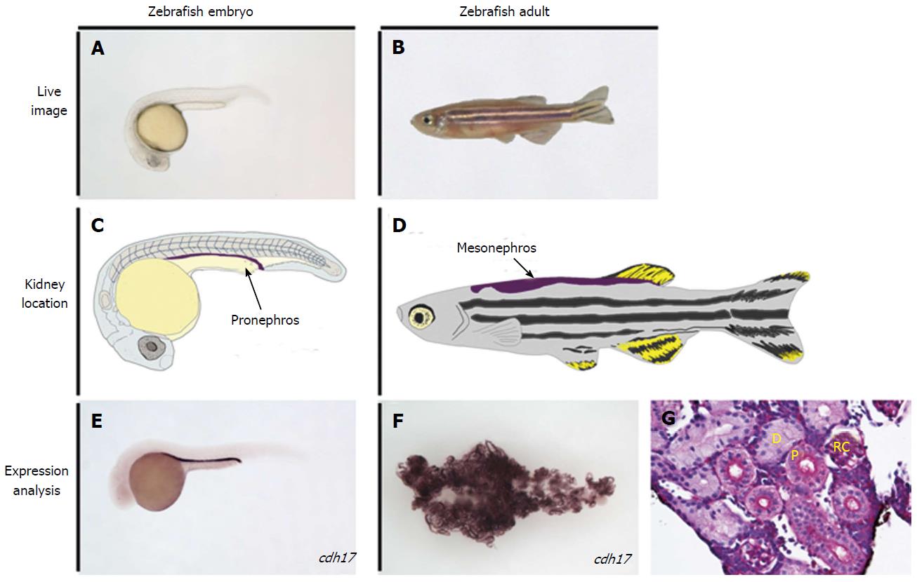
Figure 2 The zebrafish kidney in the embryo and adult.
A, B: Live image of an embryonic zebrafish at 24 h post fertilization (hpf) and at the adult stage, respectively; C: Illustration of a zebrafish at the 24 hpf stage, with the location of the embryonic kidney, the pronephros, indicated in purple; D: Illustration of an adult zebrafish with the location of the mature kidney, the mesonephros, indicated in purple; E, F: Whole mount in situ hybridization performed on the zebrafish embryo and adult kidney, respectively, to mark the expression of the tubule and duct marker gene cdh17; G: Histology of a healthy adult zebrafish kidney, stained with periodic-acid Schiff to observe nephron structures. D: Distal tubule; P: Proximal tubule; RC: Renal corpuscle; cdh17: cadherin17.
- Citation: Drummond BE, Wingert RA. Insights into kidney stem cell development and regeneration using zebrafish. World J Stem Cells 2016; 8(2): 22-31
- URL: https://www.wjgnet.com/1948-0210/full/v8/i2/22.htm
- DOI: https://dx.doi.org/10.4252/wjsc.v8.i2.22


