Copyright
©The Author(s) 2015.
World J Stem Cells. Jun 26, 2015; 7(5): 873-882
Published online Jun 26, 2015. doi: 10.4252/wjsc.v7.i5.873
Published online Jun 26, 2015. doi: 10.4252/wjsc.v7.i5.873
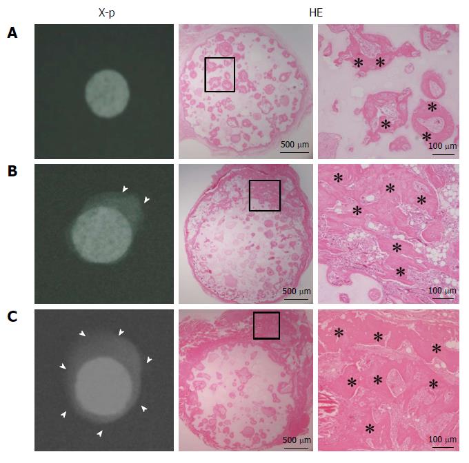
Figure 1 X-ray images and hematoxylin and eosin-stained sections at 4 wk post-implantation.
A: BMSC/TCP constructs (C group); B: Sheet/TCP constructs (S group); C: BMSC/TCP/Sheet constructs (SC group). The S and SC groups showed abundant calcification around the TCP disk. Arrowheads indicate the area of calcification. Bone formation was observed histologically in the pores of the TCP disk in all groups. The S and SC groups showed bone formation on the surface of the disk. Higher magnification images of the boxed areas are shown at the right side of each panel. Asterisks indicate bone tissue. HE: Hematoxylin and eosin.
Figure 2 ALP activity (A) and osteocalcin content (B) of harvested constructs.
The C, S, and SC groups indicate BMSC/TCP (black column), Sheet/TCP (gray column), and BMSC/TCP/Sheet (white column) constructs, respectively. Values are means ± SD (n = 6). aP < 0.05. BMSCs: Bone marrow stromal cells; TCP: Tricalcium phosphate.
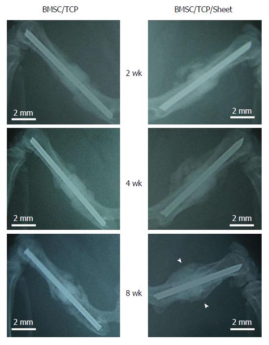
Figure 3 X-ray images of constructs implanted into femurs.
The femoral shaft was removed and replaced with BMSC/TCP constructs with or without cell sheets. Callus formation appeared around the BMSC/TCP constructs with cell sheets (BMSC/TCP/Sheet) at 2 wk post-implantation, was extended at 4 wk, and continued to the host bone at 8 wk. In contrast, the BMSC/TCP constructs showed non-union at 8 wk. Arrowheads indicate continuous bone formation. Bar = 2 mm. BMSCs: Bone marrow stromal cells; TCP: Tricalcium phosphate.
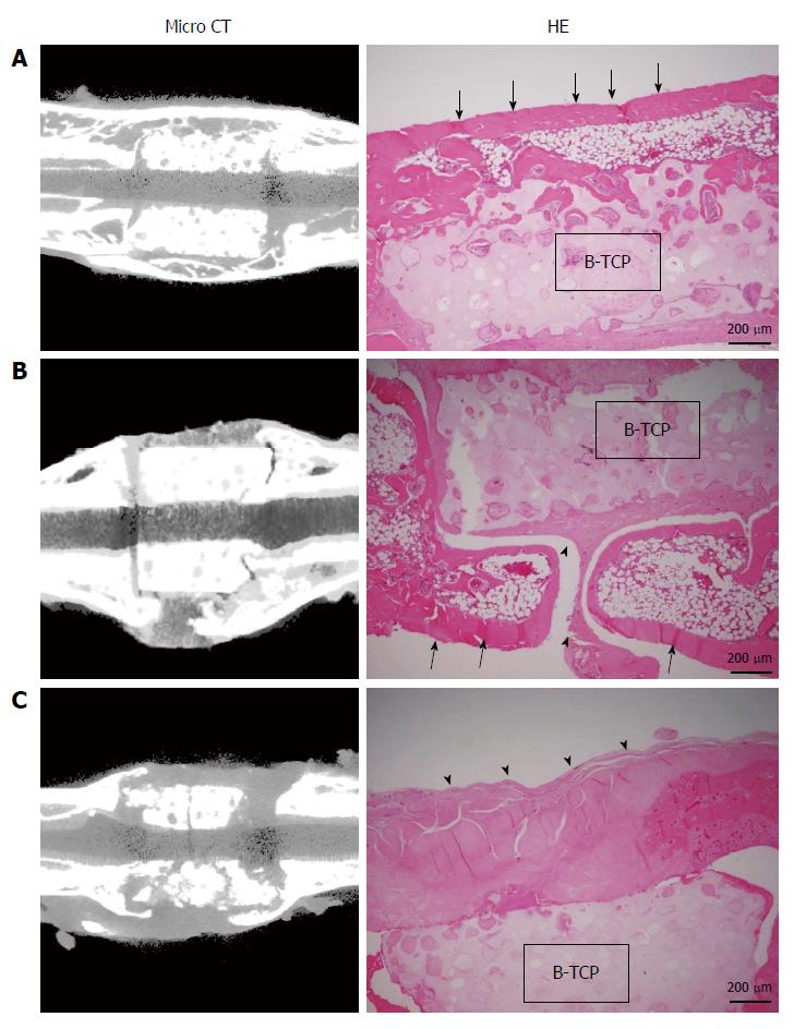
Figure 4 Micro-computed tomography images and hematoxylin and eosin-stained sections at 8 wk post-implantation.
A: BMSC/TCP/Sheet constructs with continuous bone formation to host bone. Both micro-computed tomography (CT) and histology showed bridging bone formation over the TCP to host bone (arrows); B: BMSC/TCP/Sheet constructs with segmental bone formation over the TCP. Soft tissue interposition was observed between the TCP and the callus (arrowheads); C: BMSC/TCP constructs implanted into femurs. The TCP cylinders of the BMSC/TCP constructs were broken. Arrowheads indicate soft tissue interposition between the TCP and the autologous femur. BMSCs: Bone marrow stromal cells; TCP: Tricalcium phosphate.
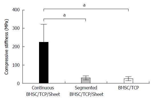
Figure 5 Results of biomechanical compression testing.
The stiffness was highest in the BMSC/TCP/Sheet constructs with continuous bone formation (black column), while the stiffness of the BMSC/TCP/Sheet constructs with segmental bone formation (gray column) was similar to that of the BMSC/TCP constructs (white column). Values are means ± SD. aP < 0.05. BMSCs: Bone marrow stromal cells; TCP: Tricalcium phosphate.
- Citation: Ueha T, Akahane M, Shimizu T, Uchihara Y, Morita Y, Nitta N, Kido A, Inagaki Y, Kawate K, Tanaka Y. Utility of tricalcium phosphate and osteogenic matrix cell sheet constructs for bone defect reconstruction. World J Stem Cells 2015; 7(5): 873-882
- URL: https://www.wjgnet.com/1948-0210/full/v7/i5/873.htm
- DOI: https://dx.doi.org/10.4252/wjsc.v7.i5.873


