Copyright
©The Author(s) 2015.
World J Stem Cells. Jun 26, 2015; 7(5): 839-851
Published online Jun 26, 2015. doi: 10.4252/wjsc.v7.i5.839
Published online Jun 26, 2015. doi: 10.4252/wjsc.v7.i5.839
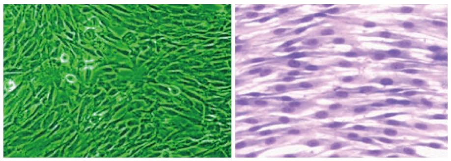
Figure 1 Phase contrast and Giemsa stained picture of Dental pulp stem cells growing in monolayer culture (Dr.
Potdar’s, laboratory).
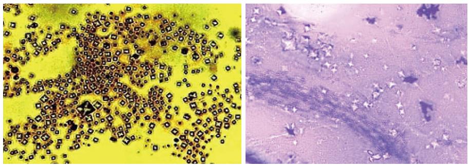
Figure 2 Silver and Giemsa stained Calcium Phosphate crystals secreted by Dental pulp stem cells in culture (Dr.
Potdar’s Laboratory).
Figure 3 Differentiation of Dental pulp stem cells into neuron like cells and cardiomyocytes (Dr.
Potdar’s Laboratory). DPSCs: Dental pulp stem cells.
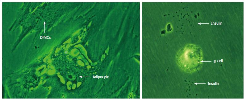
Figure 4 Differentiation of Dental pulp stem cells into adipocytes and insulin secreting beta cells.
DPSCs: Dental pulp stem cells.
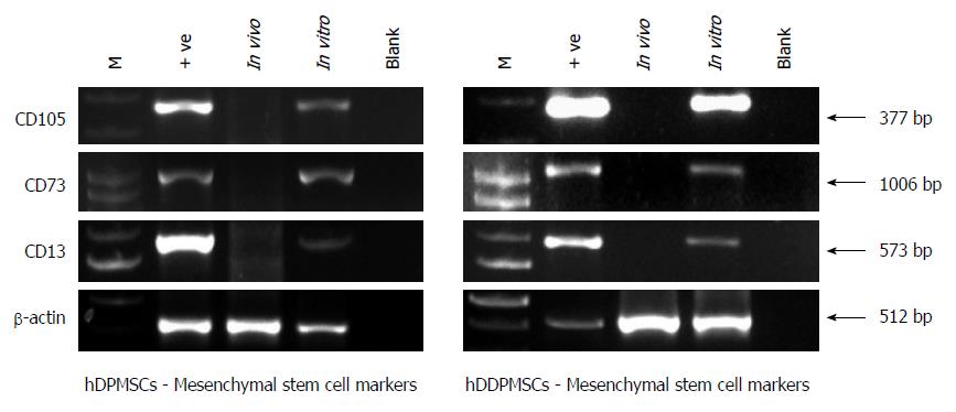
Figure 5 Mesenchymal stem cell markers in dental pulp stem cells.
- Citation: Potdar PD, Jethmalani YD. Human dental pulp stem cells: Applications in future regenerative medicine. World J Stem Cells 2015; 7(5): 839-851
- URL: https://www.wjgnet.com/1948-0210/full/v7/i5/839.htm
- DOI: https://dx.doi.org/10.4252/wjsc.v7.i5.839


