Copyright
©The Author(s) 2015.
World J Stem Cells. Jan 26, 2015; 7(1): 51-64
Published online Jan 26, 2015. doi: 10.4252/wjsc.v7.i1.51
Published online Jan 26, 2015. doi: 10.4252/wjsc.v7.i1.51
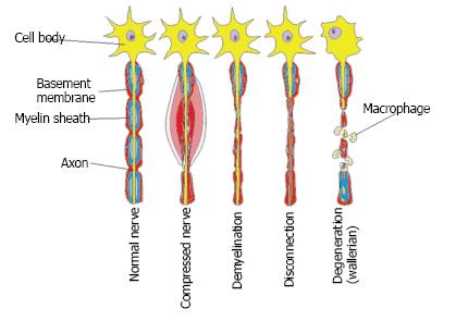
Figure 1 Changes occurring in a peripheral nerve following compression injuries adopted and modified from Ref[96].
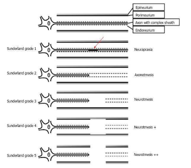
Figure 2 Sunderland classification of nerve injury adapted from Deumens et al[9], 2010.
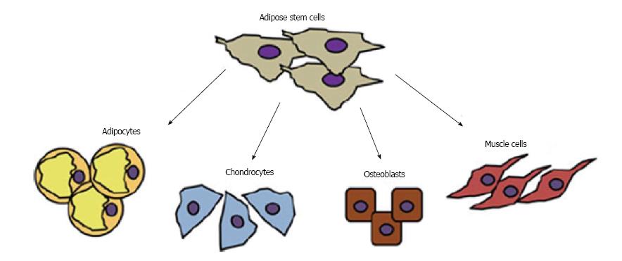
Figure 3 Differentiation of adipose derived stem cells into adipocytes, chondrocytes, osteoblasts and muscle cells adapted from Minteer et al[97].
Figure 4 Morphological differences in stimulated vs unstimulated adipose derived stem cell.
A: Unstimulated adipose derived stem cells (ADSCs) grown under normal conditions with fibroblast like morphology; B and C: Induced ADSC with a neuronal like phenotype reproduced from Cardozo et al[40] 2012. hASCs: Human adipose-derived stem cells.
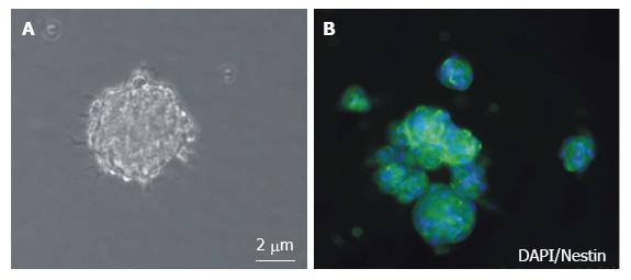
Figure 5 Appearance of neurospheres (A) Phase contrast image of free floating neurospheres; B: Fluorescence staining of nestin (Green) and nuclear stain (Blue) of neurosphere adapted from Radtke et al[43], 2009.
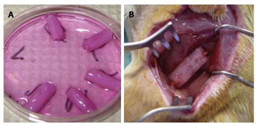
Figure 6 Fibrin nerve conduits.
A: Fibrin conduits placed in DMEM prior to seeding with ADSCs; B: Fibrin conduit being inserted surgically to bridge a rat sciatic nerve injury[42]. ADSCs: Adipose derived stem cells; DMEM: Dulbecco's modified eagle medium.
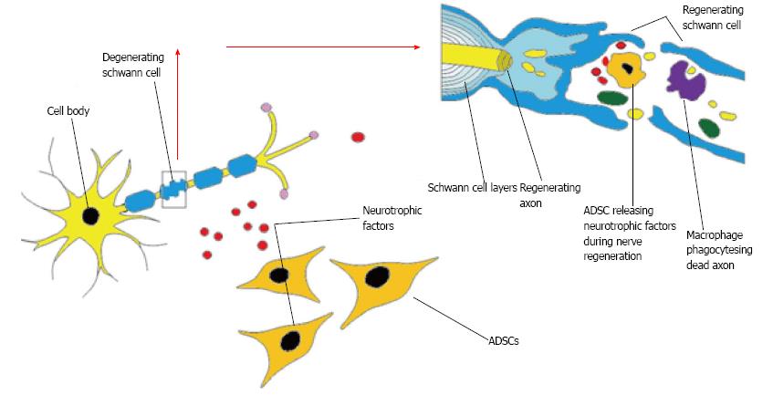
Figure 7 Potential interaction of adipose derived stem cells and the effects on peripheral nerve regeneration.
ADSCs: Adipose derived stem cells.
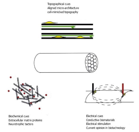
Figure 8 Interactions of topography, biochemical and electrical cues in engineering nerve tissues taken from[72].
- Citation: Zack-Williams SD, Butler PE, Kalaskar DM. Current progress in use of adipose derived stem cells in peripheral nerve regeneration. World J Stem Cells 2015; 7(1): 51-64
- URL: https://www.wjgnet.com/1948-0210/full/v7/i1/51.htm
- DOI: https://dx.doi.org/10.4252/wjsc.v7.i1.51


