Copyright
©2014 Baishideng Publishing Group Co.
World J Stem Cells. Apr 26, 2014; 6(2): 82-93
Published online Apr 26, 2014. doi: 10.4252/wjsc.v6.i2.82
Published online Apr 26, 2014. doi: 10.4252/wjsc.v6.i2.82
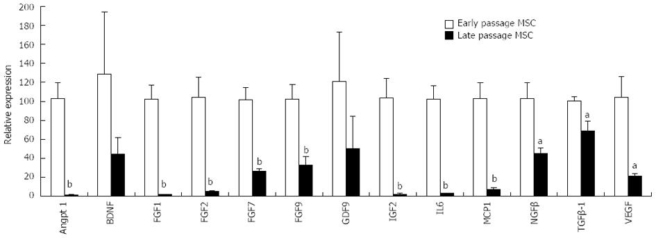
Figure 1 Ex vivo expansion of mesenchymal stem cells reduces expression of growth factor/cytokine genes.
Porcine mesenchymal stem cells (MSCs) were expanded as described[42]. Threshold cycle (CT) for the illustrated genes was determined by real-time reverse transcription polymerase chain reaction. Early and late passage MSCs received less than 5 and more than 10 trypsin passages, respectively. aP < 0.05, bP < 0.01 vs arly passage MSCs.
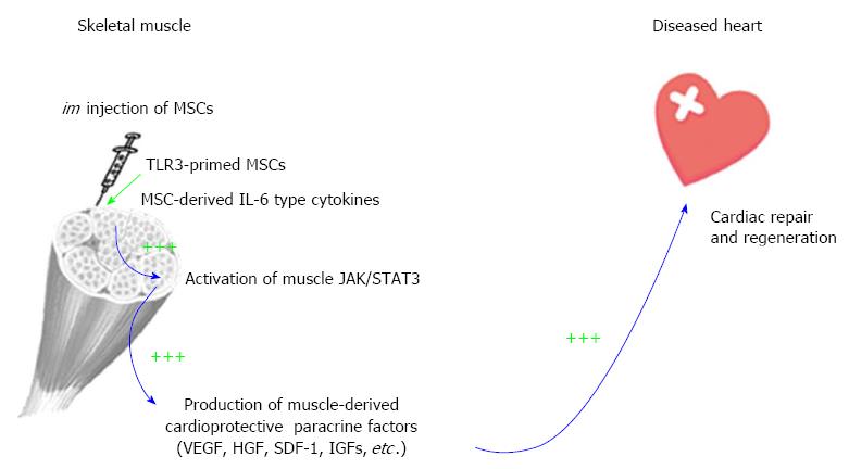
Figure 2 Intramuscular administration of mesenchymal stem cells mediates a paracrine mechanism of distal organ repair.
The paracrine cascade initiated by mesenchymal stem cells (MSCs) is illustrated by blue arrows. TLR3 priming by poly (I:C) generates a super MSC phenotype through amplification of paracrine factors, which enhances MSC potency for cardiac repair (indicated by triple green plus signs). Supporting data have been published[6,30,31,45,126].
- Citation: Mastri M, Lin H, Lee T. Enhancing the efficacy of mesenchymal stem cell therapy. World J Stem Cells 2014; 6(2): 82-93
- URL: https://www.wjgnet.com/1948-0210/full/v6/i2/82.htm
- DOI: https://dx.doi.org/10.4252/wjsc.v6.i2.82


