Copyright
©2014 Baishideng Publishing Group Co.
World J Stem Cells. Jan 26, 2014; 6(1): 11-23
Published online Jan 26, 2014. doi: 10.4252/wjsc.v6.i1.11
Published online Jan 26, 2014. doi: 10.4252/wjsc.v6.i1.11
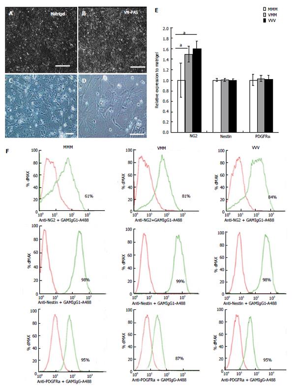
Figure 1 Oligodendrocyte progenitor cells derived from human pluripotent stem cells.
A: Morphology of day 41 Oligodendrocyte progenitor cells (OPCs) derived from cells grown on Matrigel; B: Morphology of day 41 OPCs derived from cells grown on vitronectin-derived synthetic peptide acrylate surface (VN-PAS); scale bar: 200 μm; C and D: Oligodendroglial morphology after OPC maturation; C: Low magnification; D: High magnification, scale bar: 100 μm; E: OPC marker expression; MMM: all the steps of human pluripotent stem cell (hPSC) expansion and differentiation were performed on Matrigel; MVV: hPSC expansion on Matrigel and differentiation on VN-PAS; VVV: All the steps of hPSC expansion and differentiation were performed on VN-PAS. aP < 0.05 vs MMM. F: Flow cytometry histograms of OPC markers; PDGFRα: Platelet-derived growth factor receptor alpha. This figure is adapted from Li et al[13].
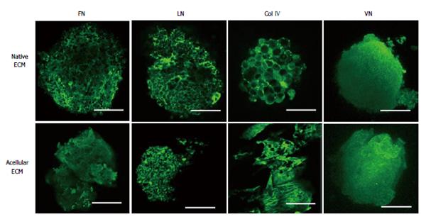
Figure 2 Three-dimensional extracellular matrix scaffolds derived from pluripotent stem cell aggregates.
Confocal images of fibronectin (FN), laminin (LN), Collagen IV (Col IV), and vitronectin (VN) expression pre- and post-decellularization [acellular extracellular matrix (ECM) and native ECM, respectively]. Scale bar: 100 μm. For native Col IV, scale bar: 50 μm. The ECM scaffolds can be used for neural differentiation. Images are adapted from Sart et al[60].
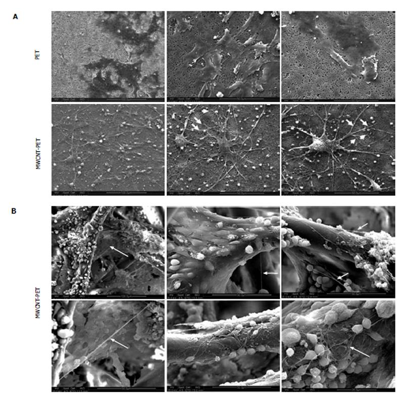
Figure 3 Neural differentiation of pluripotent stem cells.
A: Neural cells derived from murine embryonic stem cells (mESCs) cultured on 2-D PET surface with or without multiwalled carbon nanotube (MWCNT) coating; B: Neural cells derived from mESCs cultured in 3-D PET scaffolds with MWCNT coating. Arrows point to neurite fibers. Images are adapted from Zang et al[83].
- Citation: Li Y, Liu M, Yan Y, Yang ST. Neural differentiation from pluripotent stem cells: The role of natural and synthetic extracellular matrix. World J Stem Cells 2014; 6(1): 11-23
- URL: https://www.wjgnet.com/1948-0210/full/v6/i1/11.htm
- DOI: https://dx.doi.org/10.4252/wjsc.v6.i1.11


