Copyright
©2012 Baishideng.
World J Stem Cells. May 26, 2012; 4(5): 35-43
Published online May 26, 2012. doi: 10.4252/wjsc.v4.i5.35
Published online May 26, 2012. doi: 10.4252/wjsc.v4.i5.35
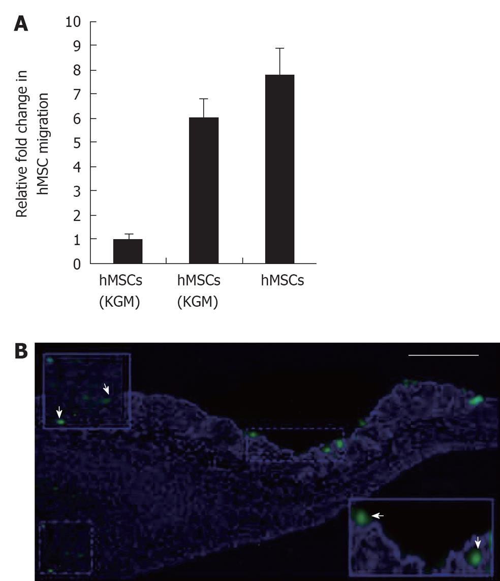
Figure 1 Migration of human mesenchymal stem cells in vitro and in vivo.
A: Human mesenchymal stem cells (HMSCs) were found to migrate toward keratinocytes as well as to KCM in greater numbers than to control medium using transwell chamber migration assay; B: Migration of locally transplanted hMSCs to the injury site. hMSCs were labeled (CFDASE) and injected at the periphery of wounded skin subcutaneously. HMSCs (green and arrows in inset) were found to migrate to the injury site (48 h post injection), scale bar 100 μm.
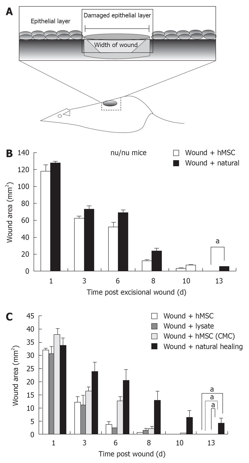
Figure 2 Excisional wound healing using human mesenchymal stem cells injection.
A: Schematic representation of excisional wound axes and the area of interest excised; B: Excisional wound was made aseptically and hMSCs (P < 0.0001, n = 5) were transplanted locally and observed on days 1, 3, 6, 8, 10 and 13; Bar graph represents wound closure over time compared with natural wound closure. Accelerated wound healing by human mesenchymal stem cells (hMSCs), hMSC lysate and concentrated conditioned medium from hMSC (CMC) in nu/nude mice; C: Macroscopic observation and bar graph representation of hMSC (P < 0.0001, n = 5), hMSC lysate (P < 0.0001, n = 5) and hMSC (CMC) (P < 0.0001, n = 5) injected wounds compared with naturally healing group after 1, 3, 6 8 10 and 13 d in nude mice. aIndicates statistically significant difference. Natural healing is essentially completed on day 14 (not shown).
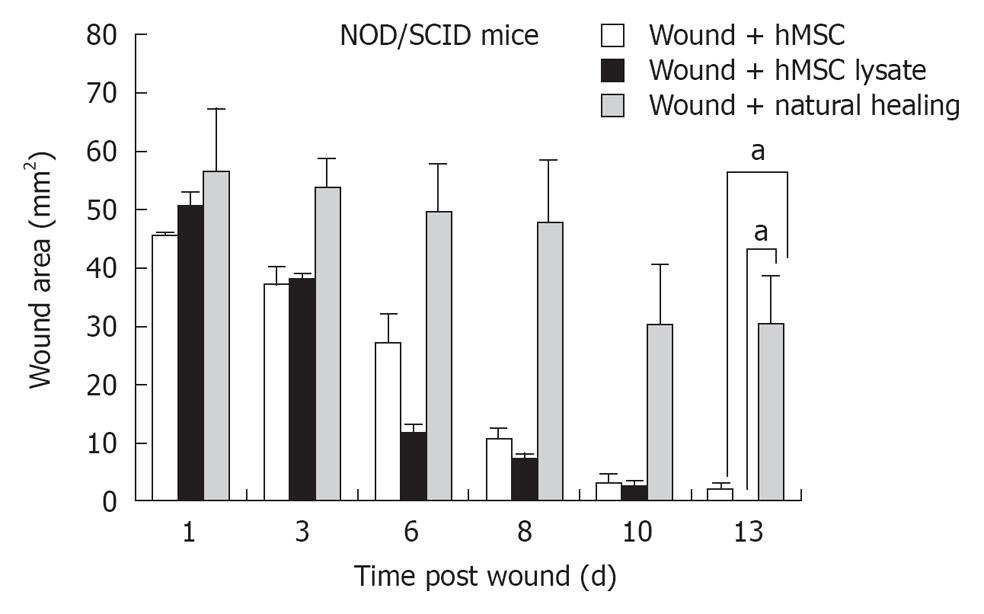
Figure 3 Accelerated wound healing by human mesenchymal stem cells and human mesenchymal stem cells lysate in a chronic wound model in NOD-SCID mice.
Graphical representation of NOD-SCID mice injected with human mesenchymal stem cells (hMSC) (P≤ 0.0003; n = 8) and hMSC lysate (P < 0.0001; n = 8), observed for wound closure after 1, 3, 6, 8,10 and 13 d and compared with naturally healing group. aIndicates statistically significant difference. Natural healing in the NOD/SCID model was completed in 24 d (not shown).
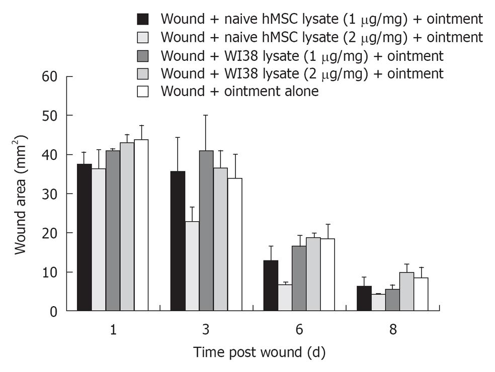
Figure 4 Emulsion containing lysate from human mesenchymal stem cells contribute significantly to wound healing.
Topical application of emulsion containing human mesenchymal stem cells (hMSCs) lysate (1-2 μg/mg) resulted in early wound closure compared with WI38 lysate (1-2 μg) and ointment alone (P≤ 0.1986; n = 4) as measured on day 8.
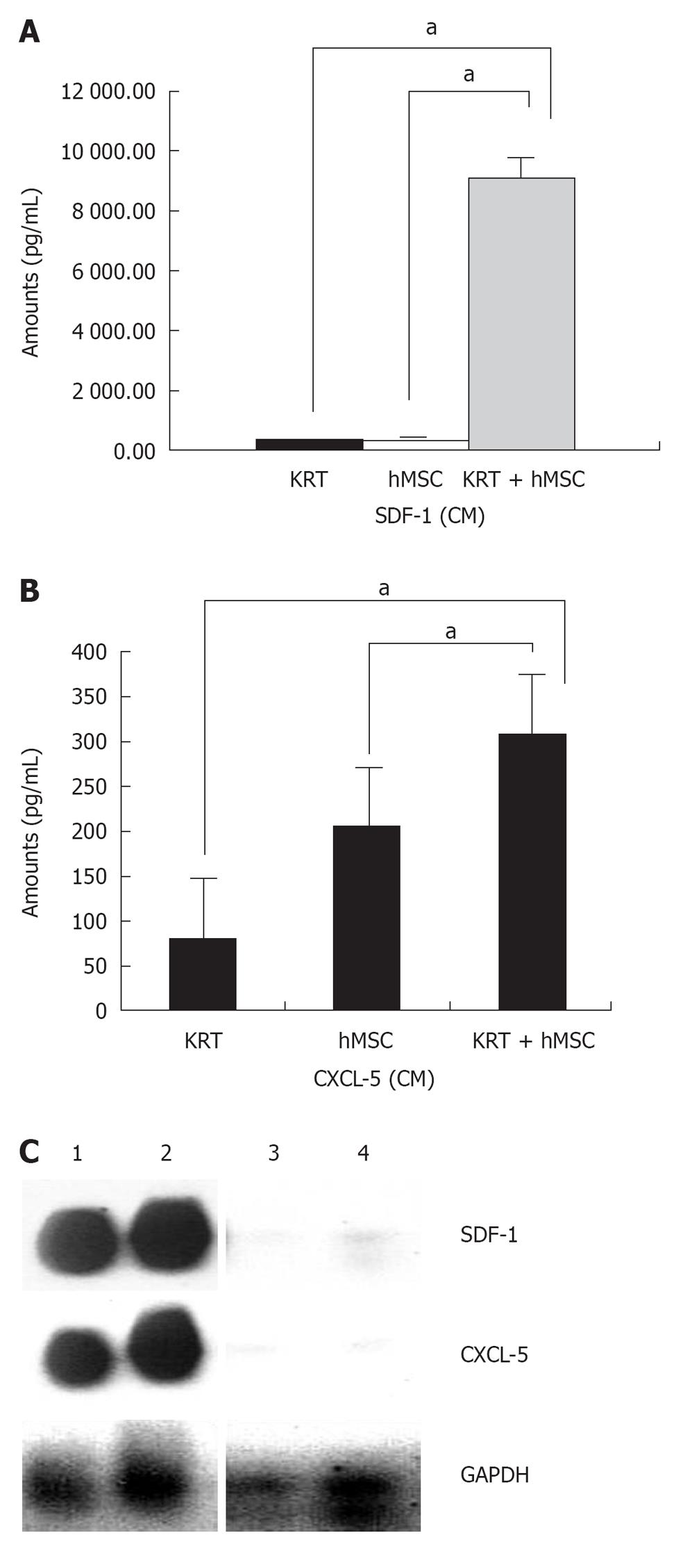
Figure 5 SDF-1 cytokine measurement in human mesenchymal stem cells co-cultured with keratinocytes.
A: SDF-1 cytokine secretion was measured in conditioned medium (CM) obtained from human mesenchymal stem cells (hMSCs) co-cultured with keratinocytes (MSC + KRT CM) compared with MSC (MSC CM) (P≤ 0.0004; n = 3) and keratinocytes (KRT CM) alone for 48 h using Multiplex assay. Increased CXCL5 (ENA-78) protein expression; B: Enzyme-linked immunosorbent assay for CXCL5 protein was performed on cell-free conditioned medium (CM) obtained from cocultured hMSCs + Keratinocytes (KRT + MSC CM) and compared with hMSC (MSC CM) alone (P≤ 0.0002; n = 3) and Keratinocytes (KRT CM) alone (P < 0.0001; n = 3) respectively. Increased expression of SDF-1 and CXCL5 was also observed in wounded skin injected with hMSC and hMSC lysate; C: SDF-1 and CXCL5 expression levels were increased in sections from hMSC and hMSC lysate injected skin (wounded) as compared with naturally healing (wounded) skin and normal skin, GAPDH was utilized as an internal control. aIndicates statistically significant difference.
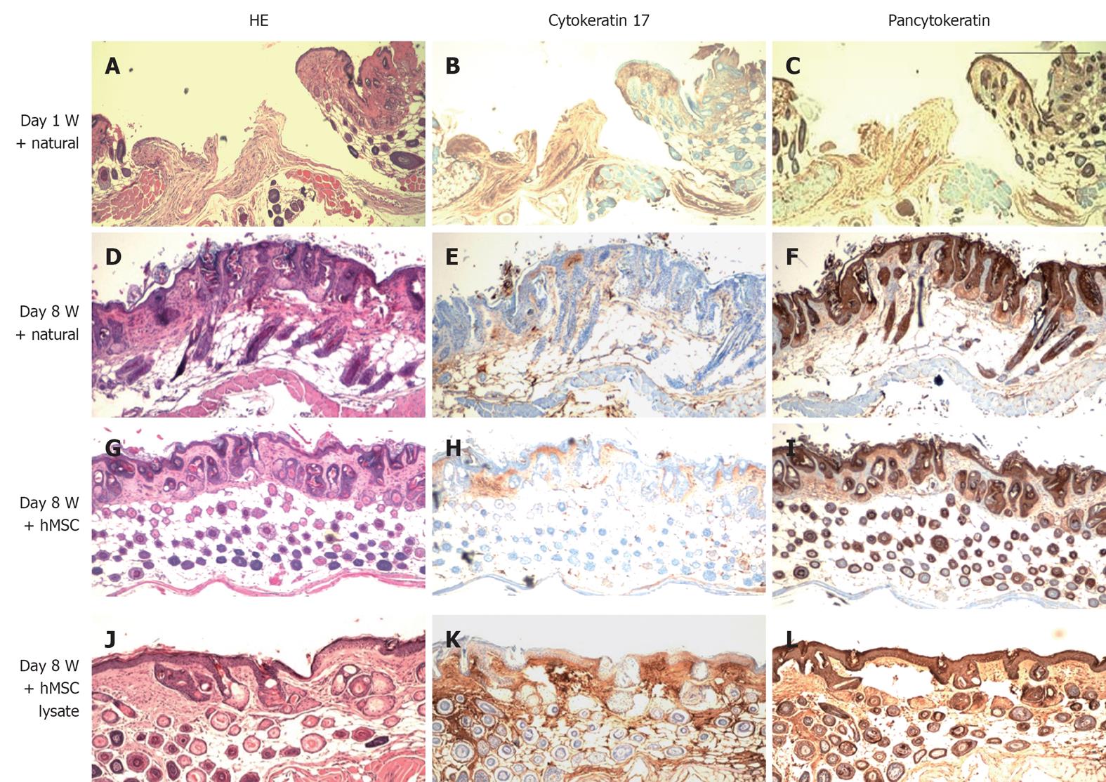
Figure 6 HE (panels A, D, G and J), cytokeratin 17 (panels B, E, H and K) and pancytokeratin staining (panels C, F, I and L) of skin sections.
Figure shows restoration of both dermis and epidermis in skins of mice (nu/nu age: 4-5 wk) treated with human mesenchymal stem cell (hMSC), hMSC lysate and CMC from hMSC; A-C: Wounded skin sections (day 1); D-F: Wounded skin allowed to heal naturally (day 8); G-I: Large numbers of pancytokeratin positive cells were observed in the dermis of hMSC administered wounded skin; J-L: hMSC Lysate injected skin sections. Scale bar: 100 μm.
- Citation: Mishra PJ, Mishra PJ, Banerjee D. Cell-free derivatives from mesenchymal stem cells are effective in wound therapy. World J Stem Cells 2012; 4(5): 35-43
- URL: https://www.wjgnet.com/1948-0210/full/v4/i5/35.htm
- DOI: https://dx.doi.org/10.4252/wjsc.v4.i5.35


