Copyright
©2012 Baishideng Publishing Group Co.
World J Stem Cells. Nov 26, 2012; 4(11): 110-116
Published online Nov 26, 2012. doi: 10.4252/wjsc.v4.i11.110
Published online Nov 26, 2012. doi: 10.4252/wjsc.v4.i11.110
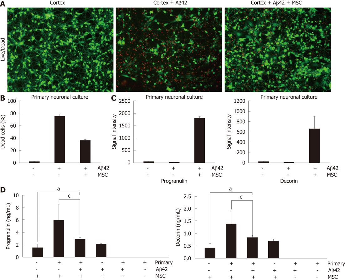
Figure 1 Decorin and progranulin are highly secreted from human umbilical cord blood-derived mesenchymal stem cells.
A: Human umbilical cord blood-derived mesenchymal stem cells (hUCB-MSCs) were co-cultured with amyloid-β42-exposed rat primary neuronal cells for 24 h in a Transwell chamber. Then, rat primary neuronal cells were stained by Live/Dead staining. Green color indicates surviving neuronal cells. B: Percentage of dead cells was calculated (P < 0.05, n = 4 per group); C: To identify paracrine factors, co-cultured media was analyzed by antibody-based array (RayBio). Spot intensity of progranulin and decorin in co-cultured media was much higher compared to rat primary neuronal cells in the absence of hUCB-MSC (P < 0.05, n = 3 per group); D: Each medium used in the Transwell was analyzed by enzyme-linked immunosorbant assay for progranulin and decorin (aP < 0.05, cP < 0.05, n = 3 per group).
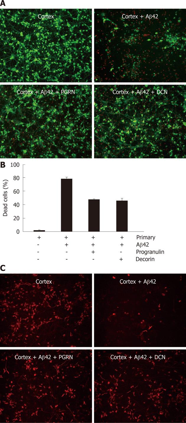
Figure 2 Recombinant progranulin and decroin protect against amyloid-β42-neurotoxicity in vitro.
A: Instead of human umbilical cord blood-derived mesenchymal stem cells, 10 ng of progranulin and decroin were used to treat amyloid-β42 (Aβ42)-exposed rat primary neuronal cells, After 36 h, each cell was assayed by Live/Dead staining. The green and red colorindicate live and dead cells, respectively; B: Percentage of dead cells in decroin- and progranulin-treated cells exposed to Aβ42; C: To show survived neurons in rat primary neuronal cells, cells were stained by anti-microtubule associated protein 2 antibody. PGRN: Progranulin; DCN: Decorin.
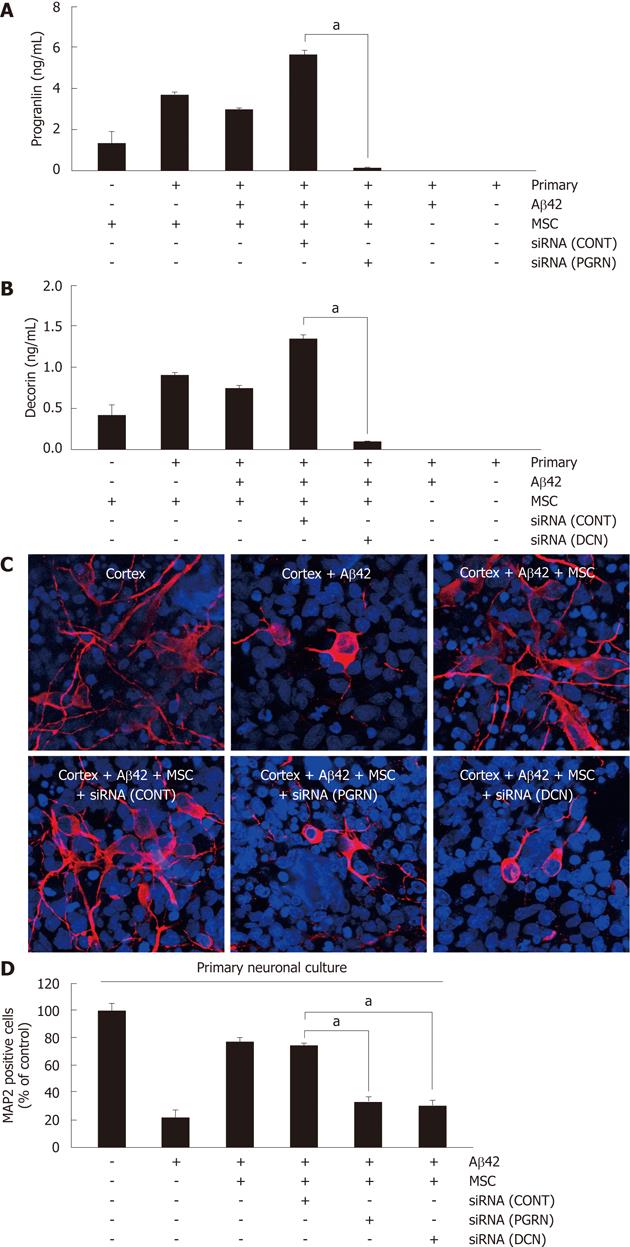
Figure 3 Knock-down of decorin and progranulin by siRNAs reduces the anti-apoptotic effect on human umbilical cord blood-derived mesenchymal stem cells in a Transwell chamber.
A, B: Each siRNA was pretreated in human umbilical cord blood-derived mesenchymal stem cells (hUCB-MSC) for 6 h. These cells were co-cultured with rat primary neuronal cells for 24 h. After co-culturing, the medium was analyzed by enzyme-linked immunosorbant assay for progranulin (A) (aP < 0.05, n = 3 per group) and decorin (B) (aP < 0.05, n = 3 per group); C: Rat primary neurons in each condition were stained by anti-microtubule associated protein 2 (MAP2) antibody. Red color indicates MAP2-positive neurons and nuclei were visualized by DAPI (blue); D: The percentage of MAP2-postive neuron in each conditions were analyzed (aP < 0.05, n = 3 per group). CONT: Control; PGRN: Progranulin; DCN: Decorin.
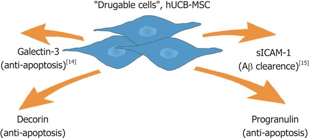
Figure 4 Paracrine action of human umbilical cord blood-derived mesenchymal stem cells in amyloid-β42 neurotoxicity in vitro.
We previously reported that human umbilical cord blood-derived mesenchymal stem cells (hUCB-MSCs) secrete soluble intercellular adhesion molecule-1 (sICAM-1) and galectin-3 for Aβ clearance and neuron survival, respectively. We also showed anti-apoptotic role of decorin and progranulin in amyloid-β42-neurotoxicity in vitro. Recently, MSCs have become regarded as drugable cells because they secrete various beneficial proteins for healing of damaged sites.
-
Citation: Kim JY, Kim DH, Kim JH, Yang YS, Oh W, Lee EH, Chang JW. Umbilical cord blood mesenchymal stem cells protect amyloid-β42 neurotoxicity
via paracrine. World J Stem Cells 2012; 4(11): 110-116 - URL: https://www.wjgnet.com/1948-0210/full/v4/i11/110.htm
- DOI: https://dx.doi.org/10.4252/wjsc.v4.i11.110


