Copyright
©2009 Baishideng.
World J Stem Cells. Dec 31, 2009; 1(1): 55-66
Published online Dec 31, 2009. doi: 10.4252/wjsc.v1.i1.55
Published online Dec 31, 2009. doi: 10.4252/wjsc.v1.i1.55
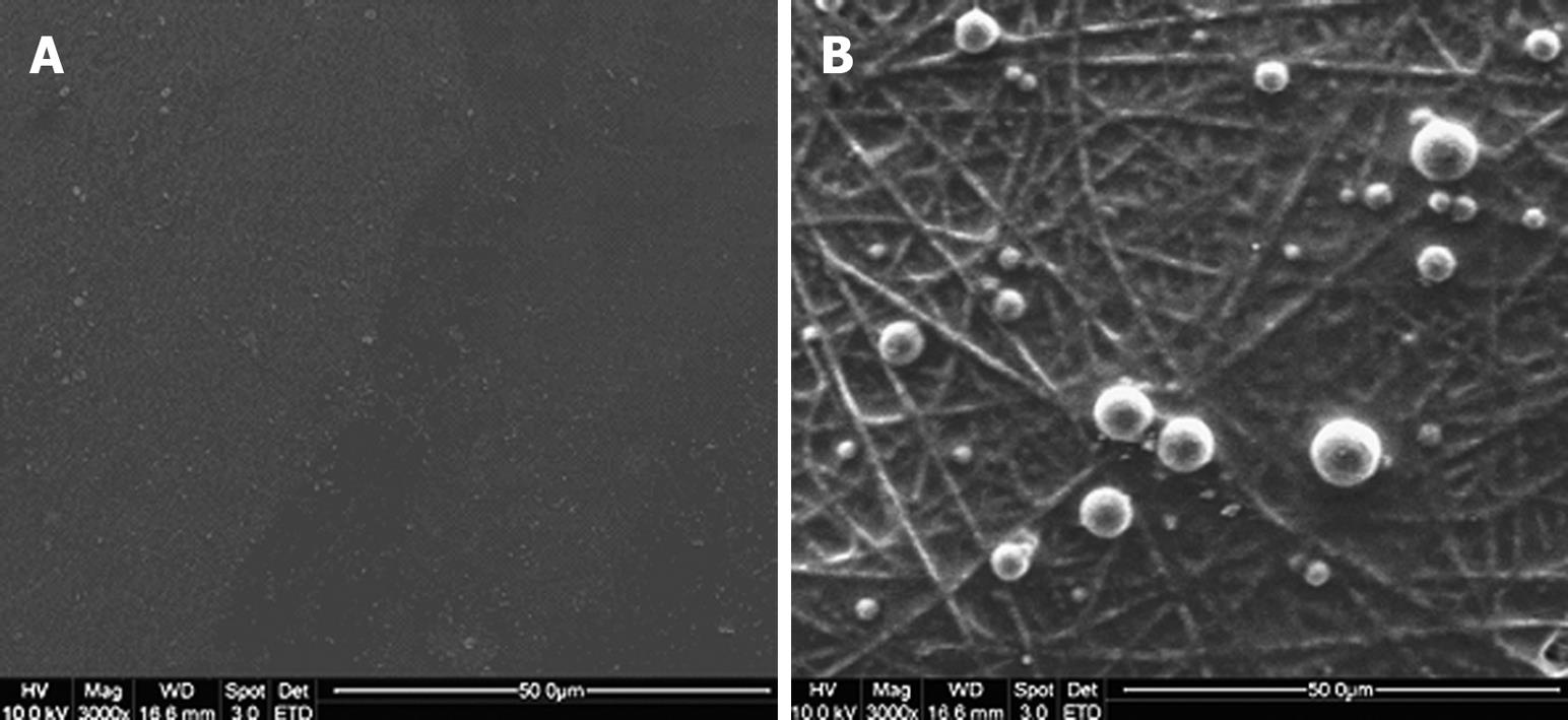
Figure 1 Capture of BM-HSCs by different substrates after 30 min of incubation.
A: No BM-HSCs captured on tissue culture polystyrene (TCP); B: Rounded morphology of BM-HSCs captured on E-selectin-coated collagen-blended polylactide-co-glycolide (PLGA) nanofiber[6].
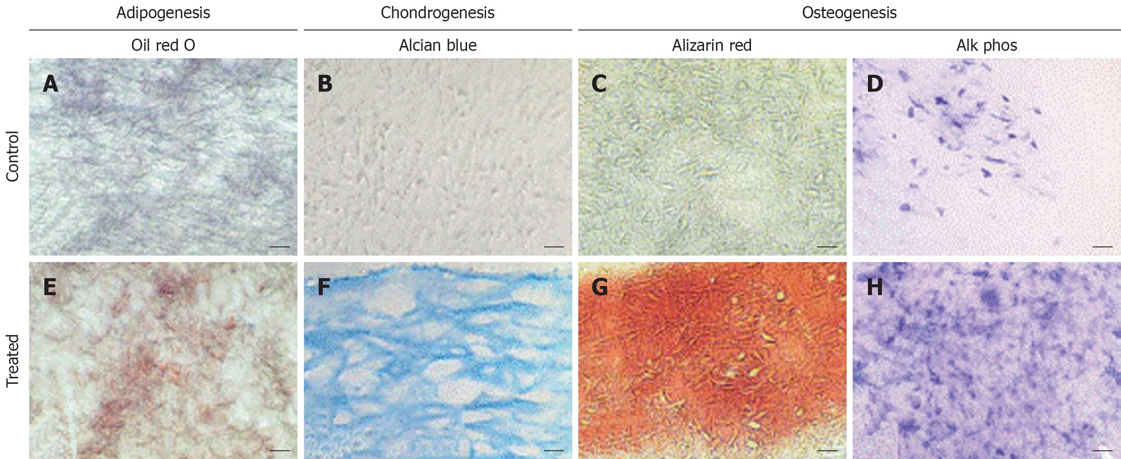
Figure 2 Histological analysis of cell-polymer constructs maintained in adipogenic, chondrogenic, or osteogenic medium.
Sections of the constructs were stained with (A,E) oil red O, (B,F) alcian blue, or (C,G) alizarin red, or histochemically stained for alkaline phosphatase (D,H). In adipogenic cultures (E), oil red O-positive lipid droplets were seen, compared to the lack of staining in the control culture (A). In chondrogenic cultures (F), intense alcian blue was seen, showing cells surrounded by a sulfated proteoglycan-rich ECM (F), whereas control cultures (B) showed little staining. In osteogenic cultures (G,H), mineralization (G) and alkaline phosphatase activity (H) were both significantly higher than in control cultures (C,D). Bar: 20 μm (B,F), 40 μm (A,C,E,G), or 80 μm (D,H)[59].
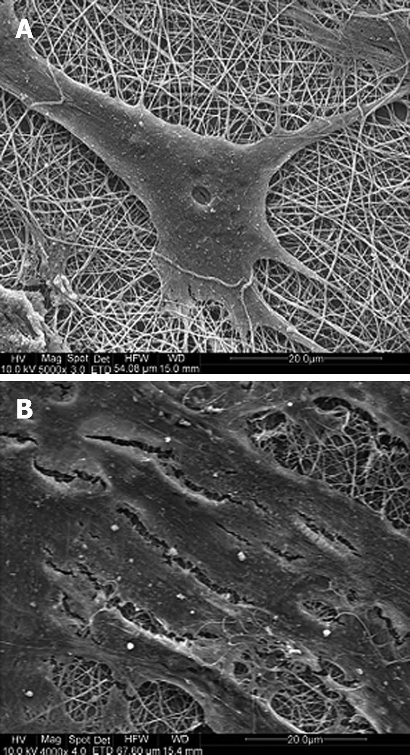
Figure 3 SEM images of (A) MSCs induced to neuronal cells grown using neuronal induction medium and (B) undifferentiated MSCs on electrospun PLA-CL/Collagen nanofibers grown using MSC growth medium[93].
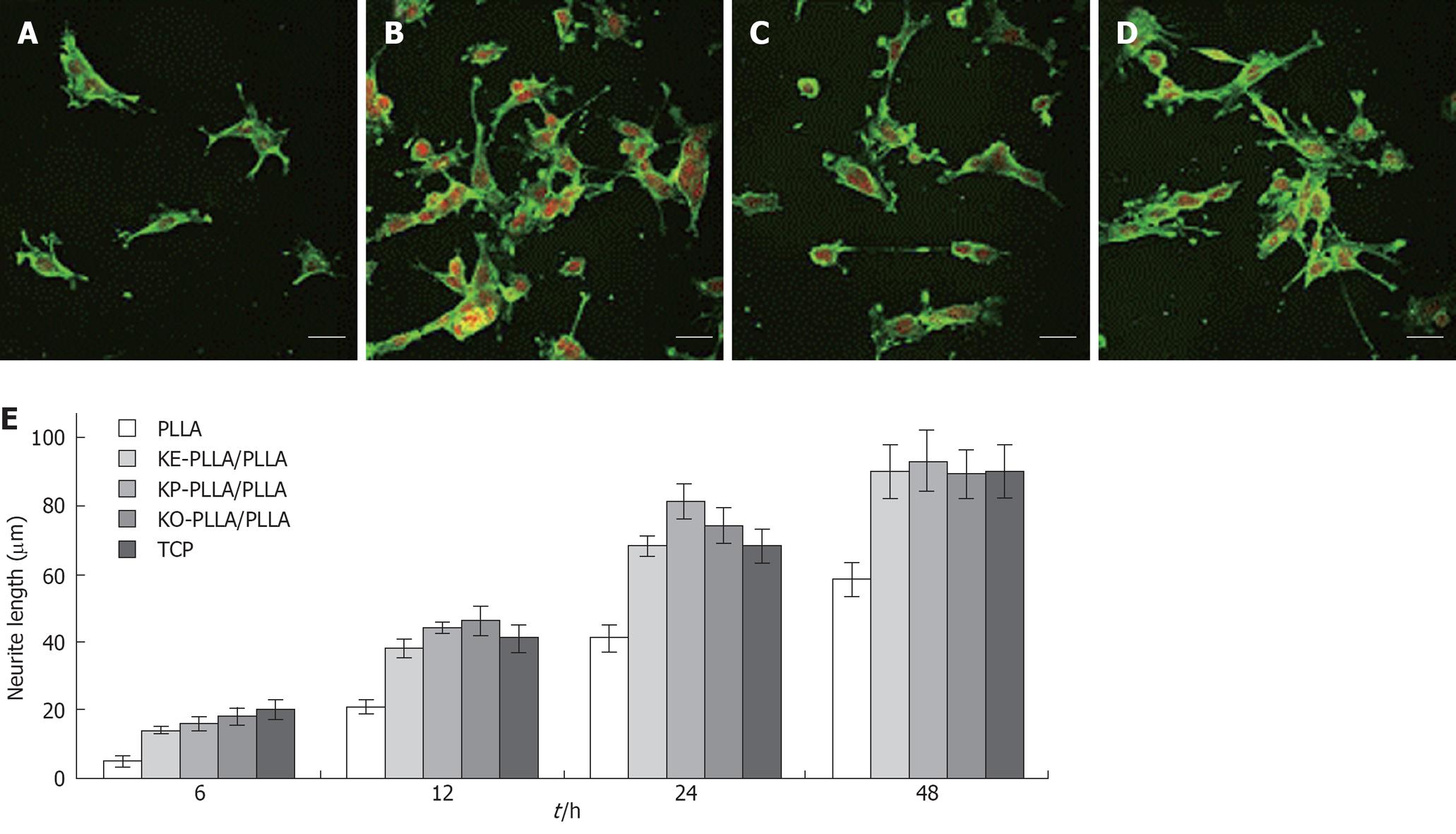
Figure 4 Laser scanning confocal microscopy (LSCM) micrographs of immunostained neurofilament 200 kDa in C17.
2 after 24 h of culture on different films. A: Poly-L-lactide acid (PLLA); B: KE-PLLA/PLLA; C: KP-PLLA/PLLA; D: KO-PLLA/PLLA; E: The average length of the longest neurite per cell from 50 randomly selected cells on different films from PLLA and K-(CH2)n-PLLA/PLLA (10/90, w/w) over cultivation[101]. The neurite was stained by FITC and nuclei was stained by PI. Scale bar: 40 μm.
- Citation: Ravichandran R, Liao S, Ng CC, Chan CK, Raghunath M, Ramakrishna S. Effects of nanotopography on stem cell phenotypes. World J Stem Cells 2009; 1(1): 55-66
- URL: https://www.wjgnet.com/1948-0210/full/v1/i1/55.htm
- DOI: https://dx.doi.org/10.4252/wjsc.v1.i1.55


