修回日期: 2022-06-17
接受日期: 2022-08-18
在线出版日期: 2022-08-28
特发性非肝硬化门脉高压症(idiopathic non-cirrhotic portal hypertension, INCPH)是一种病因不明的肝门窦血管性疾病. 临床以门脉高压的症状和体征, 而无肝硬化为特征, 初次误诊漏诊率极高. 肝活检为明确诊断的唯一途径. 近年来, CT影像等非侵入性诊断INCPH成为关注的热点. 本文归纳了INCPH的病理、CT/MRI特征以及与肝硬化的鉴别要点, 旨在提高临床医生对INCPH的认识, 减少初次诊断的误诊漏诊率.
核心提要: 特发性非肝硬化门脉高压症(idiopathic non-cirrhotic portal hypertension, INCPH)临床容易误诊和漏诊, 肝活检病理诊断仍是INCPH的金标准. 在临床实践中, 首先要根据相关指南掌握肝硬化的诊断要点, 而CT/MRI的影像特征在INCPH早期诊断及与肝硬化的鉴别中具有了较重要价值.
引文著录: 谭明洁, 刘晖, 丁惠国. 特发性非肝硬化门脉高压症的肝脏病理及影像特征. 世界华人消化杂志 2022; 30(16): 729-734
Revised: June 17, 2022
Accepted: August 18, 2022
Published online: August 28, 2022
Idiopathic non-cirrhotic portal hypertension (INCPH), a kind of portal sinus vascular disease with unknown etiology, is characterized by the presence of clinical signs and symptoms of portal hypertension (PH) in the absence of liver cirrhosis or known risk factors accountable for PH. It has an extremely high rate of initial misdiagnosis and underdiagnosis. Liver biopsy is the only way to make a definitive diagnosis. Non-invasive modalities, such as CT imaging, are becoming a hot topic of interest in recent years. This article summarizes the pathological and CT/MRI features of INCPH and the key points of differentiation from cirrhosis, to improve clinicians' understanding of INCPH and reduce the rate of initial misdiagnosis and missed diagnoses.
- Citation: Tan MJ, Liu H, Ding HG. Pathological and imaging features of idiopathic non-cirrhotic portal hypertension. Shijie Huaren Xiaohua Zazhi 2022; 30(16): 729-734
- URL: https://www.wjgnet.com/1009-3079/full/v30/i16/729.htm
- DOI: https://dx.doi.org/10.11569/wcjd.v30.i16.729
临床上大约90%的门脉高压症(portal hypertension, PH)由肝硬化引起[1], 由于专业知识的匮乏和经验不足, 非肝硬化性门脉高压症(non-cirrhotic portal hypertension, NCPH)常被临床医生误诊或漏诊. 特发性非肝硬化门脉高压症(idiopathic non-cirrhotic portal hypertension, INCPH)是在排除肝硬化和其他已知原因非肝硬化门脉高压症的情况下, 以门脉高压的症状与体征为临床特征的原因不明的肝门窦血管异常(porto-sinusoidal vascular disease, PSVD). 肝活检是INCPH诊断的唯一方法. 近年, 增强电子计算机断层扫描影像(computed tomography imaging, CT)和核磁共振成像(magnetic resonance imaging, MRI)显示了对INCPH较好的诊断价值[2], 为INCPH的早期诊断和鉴别诊断提供了新的希望. 为此, 本文概述了INCPH的病理、CT/MRI特征以及与肝硬化的鉴别要点, 旨在提高临床医生对INCPH的认识, 减少初次诊断的误诊漏诊率.
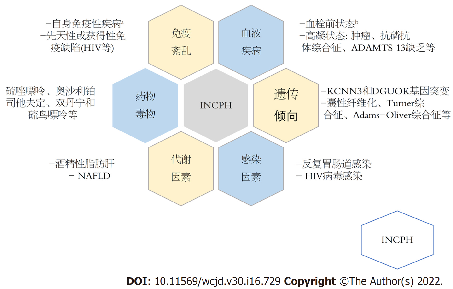
INCPH在不同国家和地区的发病率差别较大, 在印度估计高达23%以上, 而西方国家估计3%-5%, 全球患病率仍不清楚[3]. INCPH的病因和发病机制尚不明确, 目前认为可能归因于肝内微小门静脉的闭塞和损伤[4], 导致在窦前水平对门脉血流的阻力增加[5]. 几种引起血管损伤的全身性疾病及毒物或药物的慢性暴露, 被认为是INCPH发病相关因素(图1). 在中国, 药物似乎是INCPH的一个重要发病因素[6]. 此外, INCPH诊断的最大挑战是与肝硬化的鉴别[7]. 有研究报道[2], INCPH误诊为肝硬化的概率高达72.1%, 诊断延迟远超肝硬化(中位延迟时间: 32 mo vs 4 mo). 由于INCPH与肝硬化门脉高压症的治疗与预后存在差异, 因此, 临床医生应重视INCPH的早期鉴别诊断.
INCPH的宏观特征主要来源于肝移植及尸解标本[3]. 早期肝脏体积正常或增大, 表面光滑. 随着病程进展还可观察到肝包膜不规则, 包膜下区域实质萎缩等改变. 晚期肝脏明显萎缩, 体积减小, 表面凹凸不平, 可见小而相对均匀的浅黄褐色结节[3]. 横切面可见肝内门静脉主干血管壁增厚, 中小门静脉分支相互靠近或临近肝包膜. 肝门附近的切面可能出现轻微的结节改变[10]. 此外, 陈旧性血栓常在较大的门静脉分支中被发现, 近期的新鲜血栓反而更少见[11]. 所有的大体特征几乎均可以通过CT/MRI影像检查获得.
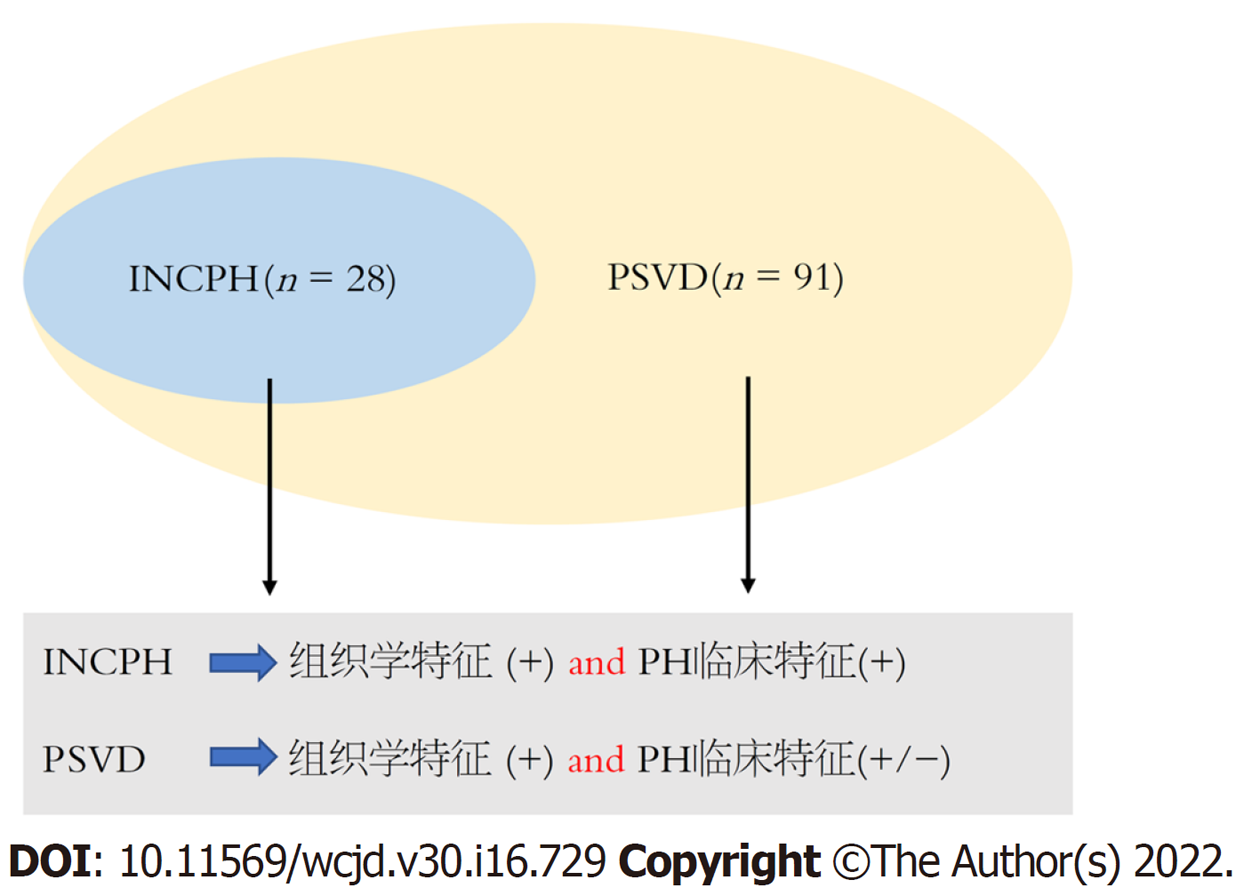
INCPH的组织学具有异质性, 在不同临床阶段, 同一疾病表现为多种病理的重叠与随机组合. 因此, 高质量的肝活检标本对INCPH的诊断非常重要. 肝活检标本必须满足: 足够长(≥20 mm)且排除肝硬化; 不能太碎(≥10个汇管区); 由病理专家阅片[8]. INCPH相对特异的三种组织学特征[8]包括: 闭塞性门静脉病(obliterative portal venopathy, OPV)、结节性再生性增生(nodular regenerative hyperplasmia, NRH)以及不完全间隔性肝硬化(incomplete septal cirrhosis, ISC). 需注意, INCPH与PSVD在自然病程上存在差异, 后者涵盖的范围更广[12], 必须区分两者的定义要素(图2).
2.2.1 OPV: OPV是INCPH唯一强有力的独立组织学预测因子[13], 被认为是INCPH/PSVD的标志性特征. 其主要特点是门静脉纤维化、血管壁不规则增厚及管腔偏心性狭窄, 有时甚至完全闭塞或门静脉缺失[8]. 正常组织学变异的汇管区二联体结构可能会与门静脉缺失相混淆, 阅片时需仔细辨别. 门静脉闭塞引起代偿性血管增生, 包括汇管区周围异常血管和肝动脉血流量代偿性增加及门体侧枝形成, 如食管胃静脉曲张等. Sato等[14]研究了28例INCPH患者肝组织谷氨酰胺合成酶(glutamine synthetase, GS)的表达发现, 71.4%患者GS阳性, 提示门静脉旁侧枝分流形成.
2.2.2 NRH: NRH是由正常肝实质转化而成的小结节, 直径1 mm-3 mm, 可分布在整个肝脏或局限于单个肝段[15]. 与肝硬化相比, 因结节周围不受纤维化包绕, 因此边界不清. 网状纤维染色可观察到结节由再生性增生和外周受压萎缩的单层肝细胞板组成, 未见纤维间隔[16]. 尽管NRH在INCPH中较少见, 但它与门脉高压的形成密不可分.
2.2.3 ISC: 特征是从汇管区发出的细薄纤维隔[16], 在肝小叶内以盲端形式终止, 不与其他细隔发生桥接. 有时可见模糊的结节, 但未见肝硬化样再生结节形成[8,17]. ISC可能是汇管区纤维化进展至终末期的表现.
除上述三个特征外, 其他的非特异性病理改变对于INCPH的诊断也有一定的价值, 主要包括汇管区异常、肝窦扩张及窦周纤维化等(表1).
| 命名 | 定义 | |
| 汇管区 | 门静脉疝 | 与汇管区周围实质直接毗邻的异常门静脉 |
| 汇管区高度血管化 | 汇管区内多个薄壁血管腔 | |
| 汇管区周围异常血管 | 位于汇管区外但紧邻汇管区的单个或多个不同口径的薄壁血管腔, 形成分流 | |
| 肝小叶 | 肝窦扩张 | 肝窦宽度大于1个肝细胞板, 通常非区域分布 |
| 窦周纤维化 | 胶原染色显示肝窦周围胶原沉积 | |
| 中央静脉异常 | 中央静脉扩张、中央静脉增厚或中央静脉多腔 |
总之, INCPH组织学异质性较大[18,19], 病理专家对INCPH的组织诊断标准也未达成一致共识, 最终诊断的确定仍需要结合临床和影像学改变.
近年来, 放射影像学研究结果在INCPH的诊断中显示了较好的应用前景.
大约71%的INCPH患者出现肝内外门静脉异常[22]. 与肝硬化的区别为: (1)门静脉通畅度: INCPH比肝硬化患者门脉通畅度低. 表现为不同程度的肝内外门静脉血栓形成、血管钙化、肝内门静脉二、三级分支的突然变细或截断及中小口径的门静脉视野不显影. INCPH患者门静脉或其主要分支异常和肝外血栓形成的概率分别为58.0%和43.0%[22], 而92.3%肝硬化患者肝内外门静脉系统均未发生闭塞[2]; (2)门静脉管壁: INCPH比肝硬化患者的门脉壁厚, 管腔直径更小. Zhao等人[23]比较了INCPH患者、肝硬化患者和健康人门静脉主干、矢状支和第3级分支的参数, 发现INCPH组和健康组的管腔直径均小于肝硬化组(P<0.05). 而相较于其他两组, INCPH组的延迟期可见门静脉系统低增强区域, 血管壁更厚, 管壁更粗糙, 门静脉主干和矢状静脉厚径比(即壁厚与管腔直径的比值)更大(P<0.05). 门静脉的异常改变可能与组织学中门脉壁增厚及纤维化有关. 因此, 对门静脉狭窄程度的定量或半定量界定, 即门静脉通畅度, 可作为INCPH严重程度的影像学分层标志.
3.2.1 尾右比异常: 80%以上的INCPH患者有肝脏形态学改变[22], 表现为尾状叶肥大和右叶萎缩. 可能归因于肝内门静脉阻塞引起灌注不足, 导致同侧节段性肝萎缩, 尾状叶代偿性肥大或灌注相对保留. 相反, 肝硬化患者更容易出现尾状叶肥大伴肝Ⅳ段的萎缩, 常用尾右比(caudate-to-right lobe ratio, C/RL), 即尾状叶与右叶的比率来描述. INCPH的尾右比明显高于肝硬化[2]. 全肝萎缩只在晚期病例中偶见, 可能代表该疾病更晚期的诊断[24,25].
3.2.2 肝脏局灶性结节性增生: 局灶性结节性增生(focal nodular hyperplasia, FNH)是INCPH较常见的表现. 这是一种由于门静脉血流量减少, 动脉血流代偿性增加导致灌注紊乱的良性增生性病变[9,26]. FNH通常单发, 典型外观为分叶状, 中央瘢痕是它的特征性表现. 钆塞酸二钠增强MRI显示, 动脉期呈高强化, 门脉期未见门脉冲洗或"快进快出"现象, 肝胆期显示周围高强度伴中央低信号瘢痕[26]. FNH在INCPH中发生率约14%, 但尚未在肝硬化患者中发现[8,27].
FNH与肝细胞性肝癌(hepatocellular carcinoma, HCC)同样都是高度血管化改变, 对于FNH在随访期间是否会发生HCC或恶性转化的结论仍不一致[2], 还需要更大的队列进行评估.
3.2.3 肝脏实质增强模式: INCPH动脉期或门脉期的异质性实质增强比肝硬化更常见. 由于门脉长期灌注不足, 肝外周动脉灌注代偿性增加, 在INCPH的动脉期或门静脉期可观察到斑片状的高信号影, 为异质性实质增强的表现. 肝实质增强程度受肝功能的显著影响[28,29]. 钆塞酸二钠增强MRI检查显示, INCPH患者肝实质信号强度比肝硬化组高[2].
3.2.4 肝包膜光滑: INCPH病变主要集中在门静脉周围[23], 因此在影像学上很少见到肝脏表面结节及肝包膜异常, 而大多数肝硬化患者都有较典型的弥漫性结节样改变[2]. INCPH肝脏表面结节的生成可能是因为远端门静脉阻塞导致局灶性肝脏表面回缩. 肝包膜结节样、不规则多出现在进展晚期, 此时与肝硬化较难辨别. 可以推测, 门脉高压表现严重, 但未见肝脏表面结节可能是INCPH与肝硬化的鉴别特征.
此外, MRI显示肝Ⅳ段萎缩导致的胆囊窝增大, 即胆囊窝扩大征, 是肝硬化的一种早期特征, 其诊断肝硬化的敏感性、特异性、准确性、阳性预测值分别为68%, 98%, 80%, 98%[30], 在INCPH的影像特征中, 也可作为一个潜在的标志物.
由于汇管区纤维化及中、小门静脉病变, INCPH的门脉高压程度明显重于肝硬化患者[2], 表现为: (1)脾脏体积明显大于肝硬化, 且多为不规则的巨脾[2,23]. 脾脏肿大还可在CT或MRI上显示多个钙化Gamna-Gandy小体, 而肝脏边缘光滑[31]; (2)食管胃底静脉曲张及出血的发生率更高. INCPH患者的死亡与静脉曲张再出血、肝性脑病及门静脉血栓形成显著相关, 几乎没有肝衰竭或肝癌的发生. 经颈静脉肝内门体静脉分流术(transjugular intrahepatic portosystemic shunt, TIPS)常用于内镜和药物治疗无效的食管胃底静脉曲张出血患者的二级预防, 但肝性脑病和血栓形成的风险增加, 尽管INCPH患者TIPS术后肝性脑病的发生率明显低于肝硬化. Bissonnette等[32]一项研究发现, 作为有TIPS指征的INCPH患者, 基础疾病的严重程度、血清肌酐浓度和腹水与不良结局相关. 因此, TIPS用于肾功能正常或无严重肝外疾病的INCPH患者. TIPS一级预防INCPH患者食管胃静脉曲张出血仍无临床研究证据.
瞬时弹性成像肝脏硬度测定(transient elastography-liver stiffness measurement, TE-LSM)被证实在INCPH的鉴别诊断中发挥良好效能. INCPH患者的肝脏硬度值正常或稍高[7.9(6.2-11.6) kPa][33], 脾脏与肝脏硬度的比值远高于肝硬化[34,35]. 在有门脉高压症状的患者中, LSM<10 kPa高度提示INCPH, 需要肝活检以确定诊断. LSM>20 kPa时INCPH的可能性非常小[33]. 有趣的是, 当INCPH患者发生NRH时, LSM值较高接近肝硬化的阈值, 此时很难将二者区分[7,36,37], 值得临床医生重视.
此外, INCPH患者的肝静脉压力梯度(hepatic venous pressure gradient, HVPG)明显低于肝硬化患者, 且与门脉高压的程度不成比例. 有研究表明[38], INCPH患者的HVPG平均为(8.3±4.5) mmHg, 约60%INCPH患者的HVPG正常或轻度升高(≤10 mmHg). 这也是与肝硬化的重要鉴别特征之一.
常见门脉高压症的表现, 而少见门脉高压症的病因, 这就是NCPH的临床特点. 迄今, 肝活检病理诊断仍是INCPH的金标准. 在临床实践中, 首先要根据相关指南, 掌握肝硬化的诊断要点, 而CT/MRI的影像特征在INCPH早期诊断及与肝硬化的鉴别中显示了较好价值. 未来, 通过多学科协作, 建立CT/MRI影像组学模型, 可能为提高INCPH的早期精准诊断带来新的希望.
学科分类: 胃肠病学和肝病学
手稿来源地: 北京市
同行评议报告学术质量分类
A级 (优秀): A, A
B级 (非常好): B
C级 (良好): C
D级 (一般): 0
E级 (差): 0
科学编辑: 张砚梁 制作编辑:张砚梁
| 2. | Kang JH, Kim DH, Kim SY, Kang HJ, Lee JB, Kim KW, Lee SS, Choi J, Lim YS. Porto-sinusoidal vascular disease with portal hypertension versus liver cirrhosis: differences in imaging features on CT and hepatobiliary contrast-enhanced MRI. Abdom Radiol (NY). 2021;46:1891-1903. [PubMed] [DOI] |
| 3. | Schouten JN, Garcia-Pagan JC, Valla DC, Janssen HL. Idiopathic noncirrhotic portal hypertension. Hepatology. 2011;54:1071-1081. [PubMed] [DOI] |
| 4. | Khanna R, Sarin SK. Non-cirrhotic portal hypertension - diagnosis and management. J Hepatol. 2014;60:421-441. [PubMed] [DOI] |
| 5. | Guido M, Sarcognato S, Sacchi D, Colloredo G. Pathology of idiopathic non-cirrhotic portal hypertension. Virchows Arch. 2018;473:23-31. [PubMed] [DOI] |
| 6. | Sun Y, Lan X, Shao C, Wang T, Yang Z. Clinical features of idiopathic portal hypertension in China: A retrospective study of 338 patients and literature review. J Gastroenterol Hepatol. 2019;34:1417-1423. [PubMed] [DOI] |
| 7. | Nicoară-Farcău O, Rusu I, Stefănescu H, Tanțău M, Badea RI, Procopeț B. Diagnostic challenges in non-cirrhotic portal hypertension - porto sinusoidal vascular disease. World J Gastroenterol. 2020;26:3000-3011. [PubMed] [DOI] |
| 8. | De Gottardi A, Rautou PE, Schouten J, Rubbia-Brandt L, Leebeek F, Trebicka J, Murad SD, Vilgrain V, Hernandez-Gea V, Nery F, Plessier A, Berzigotti A, Bioulac-Sage P, Primignani M, Semela D, Elkrief L, Bedossa P, Valla D, Garcia-Pagan JC; VALDIG group. Porto-sinusoidal vascular disease: proposal and description of a novel entity. Lancet Gastroenterol Hepatol. 2019;4:399-411. [PubMed] [DOI] |
| 9. | Kmeid M, Liu X, Ballentine S, Lee H. Idiopathic Non-Cirrhotic Portal Hypertension and Porto-Sinusoidal Vascular Disease: Review of Current Data. Gastroenterology Res. 2021;14:49-65. [PubMed] [DOI] |
| 10. | Lee H, Rehman AU, Fiel MI. Idiopathic Noncirrhotic Portal Hypertension: An Appraisal. J Pathol Transl Med. 2016;50:17-25. [PubMed] [DOI] |
| 11. | Albuquerque A, Cardoso H, Lopes J, Cipriano A, Carneiro F, Macedo G. Familial occurrence of nodular regenerative hyperplasia of the liver. Am J Gastroenterol. 2013;108:150-151. [PubMed] [DOI] |
| 12. | Wöran K, Semmler G, Jachs M, Simbrunner B, Bauer DJM, Binter T, Pomej K, Stättermayer AF, Schwabl P, Bucsics T, Paternostro R, Lampichler K, Pinter M, Trauner M, Mandorfer M, Stift J, Reiberger T, Scheiner B. Clinical Course of Porto-Sinusoidal Vascular Disease Is Distinct From Idiopathic Noncirrhotic Portal Hypertension. Clin Gastroenterol Hepatol. 2022;20:e251-e266. [PubMed] [DOI] |
| 13. | Liang J, Shi C, Dupont WD, Salaria SN, Huh WJ, Correa H, Roland JT, Perri RE, Washington MK. Key histopathologic features in idiopathic noncirrhotic portal hypertension: an interobserver agreement study and proposal for diagnostic criteria. Mod Pathol. 2021;34:592-602. [PubMed] [DOI] |
| 14. | Sato Y, Harada K, Sasaki M, Nakanuma Y. Altered intrahepatic microcirculation of idiopathic portal hypertension in relation to glutamine synthetase expression. Hepatol Res. 2015;45:1323-1330. [PubMed] [DOI] |
| 15. | Sempoux C, Balabaud C, Paradis V, Bioulac-Sage P. Hepatocellular nodules in vascular liver diseases. Virchows Arch. 2018;473:33-44. [PubMed] [DOI] |
| 17. | Aggarwal S, Fiel MI, Schiano TD. Obliterative portal venopathy: a clinical and histopathological review. Dig Dis Sci. 2013;58:2767-2776. [PubMed] [DOI] |
| 18. | Elkrief L, Rautou PE. Idiopathic non-cirrhotic portal hypertension: the tip of the obliterative portal venopathies iceberg? Liver Int. 2016;36:325-327. [PubMed] [DOI] |
| 19. | Riggio O, Gioia S, Pentassuglio I, Nicoletti V, Valente M, d'Amati G. Idiopathic noncirrhotic portal hypertension: current perspectives. Hepat Med. 2016;8:81-88. [PubMed] [DOI] |
| 20. | Schouten JN, Verheij J, Seijo S. Idiopathic non-cirrhotic portal hypertension: a review. Orphanet J Rare Dis. 2015;10:67. [PubMed] [DOI] |
| 21. | Guido M, Alves VAF, Balabaud C, Bathal PS, Bioulac-Sage P, Colombari R, Crawford JM, Dhillon AP, Ferrell LD, Gill RM, Hytiroglou P, Nakanuma Y, Paradis V, Quaglia A, Rautou PE, Theise ND, Thung S, Tsui WMS, Sempoux C, Snover D, van Leeuwen DJ; International Liver Pathology Study Group. Histology of portal vascular changes associated with idiopathic non-cirrhotic portal hypertension: nomenclature and definition. Histopathology. 2019;74:219-226. [PubMed] [DOI] |
| 22. | Glatard AS, Hillaire S, d'Assignies G, Cazals-Hatem D, Plessier A, Valla DC, Vilgrain V. Obliterative portal venopathy: findings at CT imaging. Radiology. 2012;263:741-750. [PubMed] [DOI] |
| 23. | Zhao ZL, Wei Y, Wang TL, Peng LL, Li Y, Yu MA. Imaging and Pathological Features of Idiopathic Portal Hypertension and Differential Diagnosis from Liver Cirrhosis. Sci Rep. 2020;10:2473. [PubMed] [DOI] |
| 24. | Cazals-Hatem D, Hillaire S, Rudler M, Plessier A, Paradis V, Condat B, Francoz C, Denninger MH, Durand F, Bedossa P, Valla DC. Obliterative portal venopathy: portal hypertension is not always present at diagnosis. J Hepatol. 2011;54:455-461. [PubMed] [DOI] |
| 25. | Cuadrado Lavín A, Aresti Zárate S, Delgado Tapia A, Figols Ladrón De Guevara FJ, Salcines Caviedes JR. Hepatoportal sclerosis: a cause of portal hypertension to bear in mind. Gastroenterol Hepatol. 2008;31:104. [PubMed] [DOI] |
| 26. | Yoneda N, Matsui O, Kitao A, Kozaka K, Kobayashi S, Sasaki M, Yoshida K, Inoue D, Minami T, Gabata T. Benign Hepatocellular Nodules: Hepatobiliary Phase of Gadoxetic Acid-enhanced MR Imaging Based on Molecular Background. Radiographics. 2016;36:2010-2027. [PubMed] [DOI] |
| 27. | Zuo C, Chumbalkar V, Ells PF, Bonville DJ, Lee H. Prevalence of histological features of idiopathic noncirrhotic portal hypertension in general population: a retrospective study of incidental liver biopsies. Hepatol Int. 2017;11:452-460. [PubMed] [DOI] |
| 28. | Verloh N, Haimerl M, Zeman F, Schlabeck M, Barreiros A, Loss M, Schreyer AG, Stroszczynski C, Fellner C, Wiggermann P. Assessing liver function by liver enhancement during the hepatobiliary phase with Gd-EOB-DTPA-enhanced MRI at 3 Tesla. Eur Radiol. 2014;24:1013-1019. [PubMed] [DOI] |
| 29. | Kim JY, Lee SS, Byun JH, Kim SY, Park SH, Shin YM, Lee MG. Biologic factors affecting HCC conspicuity in hepatobiliary phase imaging with liver-specific contrast agents. AJR Am J Roentgenol. 2013;201:322-331. [PubMed] [DOI] |
| 30. | Ito K, Mitchell DG, Gabata T, Hussain SM. Expanded gallbladder fossa: simple MR imaging sign of cirrhosis. Radiology. 1999;211:723-726. [PubMed] [DOI] |
| 31. | Arora A, Sarin SK. Multimodality imaging of obliterative portal venopathy: what every radiologist should know. Br J Radiol. 2015;88:20140653. [PubMed] [DOI] |
| 32. | Bissonnette J, Garcia-Pagán JC, Albillos A, Turon F, Ferreira C, Tellez L, Nault JC, Carbonell N, Cervoni JP, Abdel Rehim M, Sibert A, Bouchard L, Perreault P, Trebicka J, Trottier-Tellier F, Rautou PE, Valla DC, Plessier A. Role of the transjugular intrahepatic portosystemic shunt in the management of severe complications of portal hypertension in idiopathic noncirrhotic portal hypertension. Hepatology. 2016;64:224-231. [PubMed] [DOI] |
| 33. | Elkrief L, Lazareth M, Chevret S, Paradis V, Magaz M, Blaise L, Rubbia-Brandt L, Moga L, Durand F, Payancé A, Plessier A, Chaffaut C, Valla D, Malphettes M, Diaz A, Nault JC, Nahon P, Audureau E, Ratziu V, Castera L, Garcia Pagan JC, Ganne-Carrie N, Rautou PE; ANRS CO12 CirVir Group. Liver Stiffness by Transient Elastography to Detect Porto-Sinusoidal Vascular Liver Disease With Portal Hypertension. Hepatology. 2021;74:364-378. [PubMed] [DOI] |
| 34. | Berzigotti A. Non-invasive evaluation of portal hypertension using ultrasound elastography. J Hepatol. 2017;67:399-411. [PubMed] [DOI] |
| 35. | Seijo S, Reverter E, Miquel R, Berzigotti A, Abraldes JG, Bosch J, García-Pagán JC. Role of hepatic vein catheterisation and transient elastography in the diagnosis of idiopathic portal hypertension. Dig Liver Dis. 2012;44:855-860. [PubMed] [DOI] |
| 36. | Laharie D, Vergniol J, Bioulac-Sage P, Diris B, Poli J, Foucher J, Couzigou P, Drouillard J, de Lédinghen V. Usefulness of noninvasive tests in nodular regenerative hyperplasia of the liver. Eur J Gastroenterol Hepatol. 2010;22:487-493. [PubMed] [DOI] |
| 37. | Vuppalanchi R, Mathur K, Pyko M, Samala N, Chalasani N. Liver Stiffness Measurements in Patients with Noncirrhotic Portal Hypertension-The Devil Is in the Details. Hepatology. 2018;68:2438-2440. [PubMed] [DOI] |
| 38. | Siramolpiwat S, Seijo S, Miquel R, Berzigotti A, Garcia-Criado A, Darnell A, Turon F, Hernandez-Gea V, Bosch J, Garcia-Pagán JC. Idiopathic portal hypertension: natural history and long-term outcome. Hepatology. 2014;59:2276-2285. [PubMed] [DOI] |


