修回日期: 2015-05-18
接受日期: 2015-06-01
在线出版日期: 2015-11-08
原发性肝内胆管癌又称肝胆管细胞癌, 是起源于肝内胆管上皮细胞的一种相对少见的恶性肿瘤, 在原发性肝脏恶性肿瘤中仅次于肝细胞癌, 流行病学研究显示其发病率有逐渐增高趋势. 外科手术治疗是唯一能够获得根治的方法, 目前仍缺乏同一单位的大样本临床研究. 由于无特异性临床表现, 本病早期诊断不易, 手术切除率低, 临床死亡率较高, 预后较差. 随着医学影像学及病理诊断技术的提高, 早期诊断率增加, 本病的总体生存率有所提高, 包括非手术治疗在内的综合治疗措施在改善预后中的作用越来越重要. 本文综述近年来关于肝内胆管癌的治疗进展, 分析其治疗现状, 以更好的指导临床实践.
核心提示: 本文结合最新文献及作者临床经验阐述了原发性肝内胆管癌的诊断和治疗进展, 分析了目前的治疗现状和研究热点. 其诊断首选肝胆脾超声和肝脏磁共振成像(magnetic resonance imaging), 提出了以手术为主的综合治疗是改善肝内胆管癌预后的重要策略, 指出关于发病机制和分子靶向药物的研究有望在未来获得重要突破.
引文著录: 高正杰, 王凤山. 原发性肝内胆管癌的诊治现状. 世界华人消化杂志 2015; 23(31): 4939-4945
Revised: May 18, 2015
Accepted: June 1, 2015
Published online: November 8, 2015
Primary intrahepatic cholangiocarcinoma (ICC) is a rare malignancy arising from intrahepatic biliary epithelial cells and is also defined as cholangiohepatoma. It is the second most common primary liver malignancy after hepatocellular carcinoma. Epidemiologic research shows that the incidence rate of ICC has increased in recent years. Till now, surgical resection remains the only effective treatment to cure the disease, but single-center large-sample clinical trials are still limited. Early diagnosis of ICC is difficult due to the lack of specific clinical manifestations. The rate of resection is low, while the mortality is high and the prognosis is poor. With the development of medical imaging and pathological diagnosis technology, the early diagnosis and overall survival rates are increasing. Comprehensive therapy including non-surgical treatment plays a more and more important role in improving the prognosis. The aim of this study is to review the advances in the diagnosis and treatment of ICC in recent years.
- Citation: Gao ZJ, Wang FS. Current diagnosis and treatment of primary intrahepatic cholangiocarcinoma. Shijie Huaren Xiaohua Zazhi 2015; 23(31): 4939-4945
- URL: https://www.wjgnet.com/1009-3079/full/v23/i31/4939.htm
- DOI: https://dx.doi.org/10.11569/wcjd.v23.i31.4939
原发性肝内胆管癌是一种肝脏恶性肿瘤, 他起源于肝内胆管上皮细胞, 相对于肝细胞癌而言比较少见. 流行病学研究[1-6]显示近年来其发病率在世界范围内有升高趋势. 发病原因仍未完全明确, 多数认为与肝炎病毒感染、肝内胆管结石、肝吸虫感染、原发性硬化性胆管炎等因素有关[7-11]. 早期缺乏临床特异性表现, 大多数病例发现时已处于肿瘤晚期, 手术切除率较低, 预后较差. 近年来, 随着医学影像技术的发展, 本病的早期诊断率升高, 总体生存率有所提高[12,13]. 外科手术切除仍然是主要有效的治疗方法, 新辅助化疗、放射治疗、射频消融治疗、术后辅助光动力学治疗近年来在临床中的应用越来越多, 在改善预后中发挥着不同程度的作用. 因此, 近来强调包括手术在内的综合治疗是提高肝内胆管癌总体生存率的重要策略.
肝内胆管癌是胆管癌的一种, 约占原发性肝脏恶性肿瘤的5%-10%[14]. 流行病学显示其发病率在世界范围内有升高趋势, 澳大利亚的流行病学调查显示近30年来肝内胆管癌的发病率及死亡率增加了20%-30%[15], 而亚洲国家发病率最高的国家为泰国, 近90%的肝脏恶性肿瘤为胆管细胞癌, 中国的发病率仅次于泰国居亚洲第2位[16]. 在欧美国家该病的发病率及死亡率在部分国家略有下降, 但总体仍呈上升趋势. 该病具有种族和性别差异, 男性多于女性, 而亚洲国家的发病率高于其他国家[6,17,18]. 目前公认的致病因素有慢性病毒性肝炎、肝内胆管结石、先天性胆管扩张症、肝吸虫感染包括华支睾吸虫和麝猫后睾吸虫感染、原发性硬化性胆管炎、肝硬化以及慢性毒物损害[10,19-24], 国内以慢性乙型肝炎感染居多. 其发病的具体病理机制目前尚未明确, 有研究[8,25-28]认为与相关基因表达的上调和下降有关.
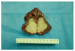
肝内胆管癌早期缺乏临床特异性表现, 与原发性肝癌临床表现相似, 大多数患者为体检发现, 多为肝内肿块产生的压迫症状, 上腹部不适、恶心等非特异性表现. 当出现胆道阻塞时可表现为梗阻性黄疸. 肿瘤晚期表现为乏力、厌食等全身消耗症状. 临床病理分型分为肿块型(59%)、管周浸润型(7%)、胆管内生长型(4%)及混合型(20%), 组织学分型以腺癌为主, 占90%以上, 其他为乳头状腺癌、印戒细胞癌等[29-31]. 肝内胆管癌患者大多发现时已处于肿瘤晚期, 临床以肿块型最为多见(图1), 发生部位上以肝左叶为多见.
3.1.1 腹部超声检查: 为肝内胆管癌患者的首选检查方法, 简便易行, 无创伤. 虽不能明确诊断, 但是可以发现肝内的占位性病变、局部扩张胆管以及有无大血管侵犯, 可清楚显示肿块内部及其周围血流分布情况. 结合动脉造影检查, 能够发现计算机断层扫描(computed tomography, CT)和磁共振成像(magnetic resonance imaging, MRI)不能发现的病灶特征[32].
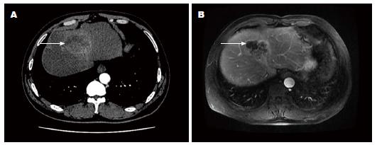
3.1.2 CT: CT在肝内胆管癌的诊断中起着重要的作用, 平扫缺乏特征性表现, 表现为肝内片状低密度影, 可见到树枝样低密度影以及扩张胆管和萎缩肝脏. 增强扫描表现为延时强化, 即"慢进慢出". 由于肿瘤缺乏血供, 增强早期, 病灶强化不明显(图2). 有助于判断肿瘤是否侵犯门脉及肝动脉、有无肝内转移以及解剖异常, 为手术治疗提供了依据和参考[33-35].
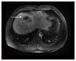
3.1.3 MRI: MRI具有多个序列成像、组织分辨率高等优点. 表现为渐进性的向心性强化(图3), 可伴有局部回缩的肝包膜、周围扩张胆管及结石. 因其组织对比性较好, 可显示小的肝内转移灶、淋巴结转移及血管侵犯[36,37]. 结合磁共振胆胰管成像检查可明确胆管异常狭窄及扩张处, 有助于定位诊断.
3.1.4 PET-CT: 正电子发射型计算机断层显像(positron emission computed tomography, PET-CT)价格昂贵, 临床应用经验较少, 主要用于术前其他检查不能明确者以及转移性肿瘤的排除诊断, 评估淋巴结的转移, 然而其发现淋巴结转移的阳性率仍不高, 临床应用有限[38-40].
糖类抗原-199(carbohydrate antigen-199, CA-199)、癌胚抗原(carcino-embryonic antigen, CEA)以及CA-125的检测在发现以及评价肝内胆管癌的预后方面具有较高的敏感性和特异性, 联合应用能够增加肝内胆管癌的诊断特异性[41]. 新的肿瘤标志物如可溶性细胞角蛋白碎片21-1(cytokeratin fragment 21-1, CYFRA21-1)、肿瘤标志物2丙氨酸激酶(tumor type M2 pyruvate kinase, TuM2-PK)以及金属蛋白酶-7(matrix metalloproteinase 7, MMP-7)能够在一定程度上帮助诊断[42]. 临床以CA-199的特异性(54%-98%)和敏感性(50%-90%)最高, 多联合其他肿瘤标志物检测, 提高诊断准确率.
目前仍然缺乏肝内胆管癌的大宗治疗经验, 缺乏单个医院的大样本临床研究. 其治疗的主要方法是外科手术切除, 尽管临床切除率不高, 仍应争取手术切除. 手术方式以规则性肝叶、肝段切除达到切缘无肿瘤残留的R0切除为首选. R0切除患者总体生存率较高, 3年生存率可达35.6%, 5年生存率可达20.1%[43,44]. 而肝移植治疗由于供体缺乏, 临床应用有限. 对于有区域淋巴结转移的患者应同时行淋巴结廓清, 或行扩大切除术, 淋巴结转移被认为是影响预后的重要因素, 但是目前关于淋巴结廓清的疗效尚有争议, 有待于进一步的临床研究[45,46]. 对于不能根治性切除患者可行化疗和放疗, 但多数学者认为本病对化疗及放疗不敏感, 但也有学者认为术后联合化疗能够改善预后[12,47,48]. 近年来非手术治疗的进展给肝内胆管癌的治疗带来了重要方法. 对于直径较小(3-5 cm)的肿瘤和不能手术根治性切除的患者也可采用射频消融治疗, 能够达到较好的效果, 并改善预后[49]. 光动力学治疗受到了越来越多的学者重视, 利用光动力效应直接杀死癌变细胞, 并可造成肿瘤血管栓塞, 引起缺血性坏死, 是一种可提高生存率的有效方法[50,51]. 对于肿瘤晚期出现胆道梗阻症状者可行姑息减黄治疗, 介入栓塞治疗, 以提高生活质量.
肝内胆管癌恶性程度高, 大多数患者就诊时已处于肿瘤晚期, 手术切除率低, 预后比肝细胞癌差. 手术切除治疗是获得长期生存的最重要因素, 本病一旦确诊, 均应行以肝切除为主的综合治疗, 预后与患者年龄、肿瘤分期、根治性切除情况、有无转移及病理类型等因素密切相关. 尽管近年来非手术治疗方法的研究取得了不少进展, 但总体预后仍不乐观. 国外报道的手术切除率为50.5%, 总体平均中位生存时间为16.1 mo, 根治性切除者可达27.6 mo, 姑息性放化疗为12.9 mo[1]. 总体手术切除率及生存率较过去有所提高, 5年生存率最高已可达31.1%[12]. 但由于本病相对少见, 各个研究涉及的病例数较少, 样本选择差异性较大, 所得结论不一. 改善预后的关键是早期诊断及治疗, 仍需要流行病学的分析, 进一步确定病因和危险因素. 同时需要进一步地关于发病机制的基础研究, 研制特异性的靶向药物, 进一步提高疗效.
肝内胆管癌是一种恶性程度较高的肝脏肿瘤, 缺乏特异性临床表现, 早期诊断不易, 远期预后较差. 早期诊断和治疗能够显著提高生存率, 临床医生应提高对该病的认识和警惕性, 对早期的非特异性消化系统症状及时行相关检查, 提高早期诊断率. 肝胆脾彩超及肝脏MRI检查为首选检查方法, 外科手术切除是唯一改善预后获得长期生存的治疗方法, 同时需结合患者的年龄、全身状况、肿瘤分期决定具体的治疗方式. 尽管近年非手术治疗方法研究有所进展, 改善预后的关键仍然是早期诊断和治疗. 相信随着医学影像技术和实验诊断技术的提高以及对本病发病机制基础研究的进步, 肝内胆管癌患者的预后会越来越好.
肝内胆管癌是一种相对少见的肝脏恶性肿瘤, 与肝细胞癌具有不同的生物学特征, 缺乏特异性临床表现, 早期诊断不易, 预后较差. 手术切除是唯一获得长期生存的治疗方法, 以手术治疗为主的综合治疗是提高本病生存率的重要策略.
薛东波, 教授, 哈尔滨医科大学附属第一医院
肝脏射频消融治疗肝内胆管癌有许多成功报道, 具有创伤小、恢复快的优点, 对直径较小的肿瘤是一种微创的治疗方法. 光动力学治疗在改善肝内胆管癌预后方面也发挥着重要的作用, 是肝内胆管癌综合治疗措施中的重要选择方法.
黄元哲等对原发性肝内胆管癌进行了相关报道, 认为其发病率近年有所升高, 早期诊断率低, 手术切除率低, 预后差, 根治性切除是改善患者预后的唯一有效途径.
本文较详细的介绍了肝内胆管癌的流行病学、诊断与治疗进展, 也介绍了肝内胆管癌的非手术治疗方法和研究热点.
本文总结了肝内胆管癌的诊治进展及当前的研究热点, 分析了目前的治疗现状, 提出了进一步研究的关键问题.
光动力学治疗: 利用光能转化过程中产生的单态氧杀死病变细胞, 同时引发毛细血管内皮损伤, 导致血管栓塞, 造成病变组织缺血性坏死, 是目前肿瘤治疗中的一种有效方法.
本文围绕肝内胆管癌比较详细地介绍了肝内胆管癌的流行病学病因学、临床病例特征、各种相关检查、常用治疗方法, 并且提出改善此病预后的方法, 对临床治疗及研究具有一定借鉴意义.
编辑: 韦元涛 电编: 都珍珍
| 1. | Dhanasekaran R, Hemming AW, Zendejas I, George T, Nelson DR, Soldevila-Pico C, Firpi RJ, Morelli G, Clark V, Cabrera R. Treatment outcomes and prognostic factors of intrahepatic cholangiocarcinoma. Oncol Rep. 2013;29:1259-1267. [PubMed] [DOI] |
| 2. | Ghouri YA, Mian I, Blechacz B. Cancer review: Cholangiocarcinoma. J Carcinog. 2015;14:1. [PubMed] [DOI] |
| 3. | Baheti AD, Tirumani SH, Rosenthal MH, Shinagare AB, Ramaiya NH. Diagnosis and management of intrahepatic cholangiocarcinoma: a comprehensive update for the radiologist. Clin Radiol. 2014;69:e463-e470. [PubMed] [DOI] |
| 4. | Tyson GL, Ilyas JA, Duan Z, Green LK, Younes M, El-Serag HB, Davila JA. Secular trends in the incidence of cholangiocarcinoma in the USA and the impact of misclassification. Dig Dis Sci. 2014;59:3103-3110. [PubMed] [DOI] |
| 5. | Njei B. Changing pattern of epidemiology in intrahepatic cholangiocarcinoma. Hepatology. 2014;60:1107-1108. [PubMed] [DOI] |
| 6. | Bertuccio P, Bosetti C, Levi F, Decarli A, Negri E, La Vecchia C. A comparison of trends in mortality from primary liver cancer and intrahepatic cholangiocarcinoma in Europe. Ann Oncol. 2013;24:1667-1674. [PubMed] [DOI] |
| 7. | Peng NF, Li LQ, Qin X, Guo Y, Peng T, Xiao KY, Chen XG, Yang YF, Su ZX, Chen B. Evaluation of risk factors and clinicopathologic features for intrahepatic cholangiocarcinoma in Southern China: a possible role of hepatitis B virus. Ann Surg Oncol. 2011;18:1258-1266. [PubMed] [DOI] |
| 8. | Subrungruanga I, Thawornkunob C, Chawalitchewinkoon-Petmitrc P, Pairojkul C, Wongkham S, Petmitrb S. Gene expression profiling of intrahepatic cholangiocarcinoma. Asian Pac J Cancer Prev. 2013;14:557-563. [PubMed] [DOI] |
| 9. | Ariizumi S, Yamamoto M. Intrahepatic cholangiocarcinoma and cholangiolocellular carcinoma in cirrhosis and chronic viral hepatitis. Surg Today. 2015;45:682-687. [PubMed] |
| 10. | Fu XH, Tang ZH, Zong M, Yang GS, Yao XP, Wu MC. Clinicopathologic features, diagnosis and surgical treatment of intrahepatic cholangiocarcinoma in 104 patients. Hepatobiliary Pancreat Dis Int. 2004;3:279-283. [PubMed] |
| 11. | Zhou YM, Yin ZF, Yang JM, Li B, Shao WY, Xu F, Wang YL, Li DQ. Risk factors for intrahepatic cholangiocarcinoma: a case-control study in China. World J Gastroenterol. 2008;14:632-635. [PubMed] [DOI] |
| 12. | Morise Z, Sugioka A, Tokoro T, Tanahashi Y, Okabe Y, Kagawa T, Takeura C. Surgery and chemotherapy for intrahepatic cholangiocarcinoma. World J Hepatol. 2010;2:58-64. [PubMed] [DOI] |
| 13. | Lubezky N, Facciuto M, Harimoto N, Schwartz ME, Florman SS. Surgical treatment of intrahepatic cholangiocarcinoma in the USA. J Hepatobiliary Pancreat Sci. 2015;22:124-130. [PubMed] [DOI] |
| 14. | Chang KY, Chang JY, Yen Y. Increasing incidence of intrahepatic cholangiocarcinoma and its relationship to chronic viral hepatitis. J Natl Compr Canc Netw. 2009;7:423-427. [PubMed] |
| 15. | Luke C, Price T, Roder D. Epidemiology of cancer of the liver and intrahepatic bile ducts in an Australian population. Asian Pac J Cancer Prev. 2010;11:1479-1485. [PubMed] |
| 16. | Shin HR, Oh JK, Masuyer E, Curado MP, Bouvard V, Fang Y, Wiangnon S, Sripa B, Hong ST. Comparison of incidence of intrahepatic and extrahepatic cholangiocarcinoma--focus on East and South-Eastern Asia. Asian Pac J Cancer Prev. 2010;11:1159-1166. [PubMed] |
| 17. | McLean L, Patel T. Racial and ethnic variations in the epidemiology of intrahepatic cholangiocarcinoma in the United States. Liver Int. 2006;26:1047-1053. [PubMed] [DOI] |
| 18. | Center MM, Jemal A. International trends in liver cancer incidence rates. Cancer Epidemiol Biomarkers Prev. 2011;20:2362-2368. [PubMed] [DOI] |
| 19. | Sriputtha S, Khuntikeo N, Promthet S, Kamsa-Ard S. Survival rate of intrahepatic cholangiocarcinoma patients after surgical treatment in Thailand. Asian Pac J Cancer Prev. 2013;14:1107-1110. [PubMed] [DOI] |
| 20. | Kim HG, Han J, Kim MH, Cho KH, Shin IH, Kim GH, Kim JS, Kim JB, Kim TN, Kim TH. Prevalence of clonorchiasis in patients with gastrointestinal disease: a Korean nationwide multicenter survey. World J Gastroenterol. 2009;15:86-94. [PubMed] [DOI] |
| 21. | Zhou Y, Zhao Y, Li B, Huang J, Wu L, Xu D, Yang J, He J. Hepatitis viruses infection and risk of intrahepatic cholangiocarcinoma: evidence from a meta-analysis. BMC Cancer. 2012;12:289. [PubMed] [DOI] |
| 22. | Uenishi T, Nagano H, Marubashi S, Hayashi M, Hirokawa F, Kaibori M, Matsui K, Kubo S. The long-term outcomes after curative resection for mass-forming intrahepatic cholangiocarcinoma associated with hepatitis C viral infection: a multicenter analysis by Osaka Hepatic Surgery Study Group. J Surg Oncol. 2014;110:176-181. [PubMed] [DOI] |
| 23. | Matsumoto K, Onoyama T, Kawata S, Takeda Y, Harada K, Ikebuchi Y, Ueki M, Miura N, Yashima K, Koda M. Hepatitis B and C virus infection is a risk factor for the development of cholangiocarcinoma. Intern Med. 2014;53:651-654. [PubMed] [DOI] |
| 24. | Trilianos P, Selaru F, Li Z, Gurakar A. Trends in pre-liver transplant screening for cholangiocarcinoma among patients with primary sclerosing cholangitis. Digestion. 2014;89:165-173. [PubMed] [DOI] |
| 25. | Grassian AR, Pagliarini R, Chiang DY. Mutations of isocitrate dehydrogenase 1 and 2 in intrahepatic cholangiocarcinoma. Curr Opin Gastroenterol. 2014;30:295-302. [PubMed] [DOI] |
| 26. | Wu WR, Zhang R, Shi XD, Zhu MS, Xu LB, Zeng H, Liu C. Notch1 is overexpressed in human intrahepatic cholangiocarcinoma and is associated with its proliferation, invasiveness and sensitivity to 5-fluorouracil in vitro. Oncol Rep. 2014;31:2515-2524. [PubMed] [DOI] |
| 27. | Yu Y, Liao M, Liu R, Chen J, Feng H, Fu Z. Overexpression of lactate dehydrogenase-A in human intrahepatic cholangiocarcinoma: its implication for treatment. World J Surg Oncol. 2014;12:78. [PubMed] [DOI] |
| 28. | Sueoka H, Hirano T, Uda Y, Iimuro Y, Yamanaka J, Fujimoto J. Blockage of CXCR2 suppresses tumor growth of intrahepatic cholangiocellular carcinoma. Surgery. 2014;155:640-649. [PubMed] [DOI] |
| 29. | Mavros MN, Economopoulos KP, Alexiou VG, Pawlik TM4. Treatment and Prognosis for Patients With Intrahepatic Cholangiocarcinoma: Systematic Review and Meta-analysis. JAMA Surg. 2014; Apr 9. [Epub ahead of print]. [PubMed] [DOI] |
| 30. | Yamasaki S. Intrahepatic cholangiocarcinoma: macroscopic type and stage classification. J Hepatobiliary Pancreat Surg. 2003;10:288-291. [PubMed] [DOI] |
| 31. | Shimada K, Sano T, Sakamoto Y, Esaki M, Kosuge T, Ojima H. Surgical outcomes of the mass-forming plus periductal infiltrating types of intrahepatic cholangiocarcinoma: a comparative study with the typical mass-forming type of intrahepatic cholangiocarcinoma. World J Surg. 2007;31:2016-2022. [PubMed] [DOI] |
| 32. | Galassi M, Iavarone M, Rossi S, Bota S, Vavassori S, Rosa L, Leoni S, Venerandi L, Marinelli S, Sangiovanni A. Patterns of appearance and risk of misdiagnosis of intrahepatic cholangiocarcinoma in cirrhosis at contrast enhanced ultrasound. Liver Int. 2013;33:771-779. [PubMed] [DOI] |
| 33. | Hua X, Fu X, Hao Z, Fu Q, Shang H, Chi D. [Computed tomographic diagnosis of intrahepatic cholangiocarcinoma]. Zhonghua Yi Xue Za Zhi. 2014;94:449-451. [PubMed] |
| 34. | Baheti AD, Tirumani SH, Shinagare AB, Rosenthal MH, Hornick JL, Ramaiya NH, Wolpin BM. Correlation of CT patterns of primary intrahepatic cholangiocarcinoma at the time of presentation with the metastatic spread and clinical outcomes: retrospective study of 92 patients. Abdom Imaging. 2014;39:1193-1201. [PubMed] [DOI] |
| 35. | Iavarone M, Piscaglia F, Vavassori S, Galassi M, Sangiovanni A, Venerandi L, Forzenigo LV, Golfieri R, Bolondi L, Colombo M. Contrast enhanced CT-scan to diagnose intrahepatic cholangiocarcinoma in patients with cirrhosis. J Hepatol. 2013;58:1188-1193. [PubMed] [DOI] |
| 36. | Sheng RF, Zeng MS, Rao SX, Ji Y, Chen LL. MRI of small intrahepatic mass-forming cholangiocarcinoma and atypical small hepatocellular carcinoma (≤3 cm) with cirrhosis and chronic viral hepatitis: a comparative study. Clin Imaging. 2014;38:265-272. [PubMed] [DOI] |
| 37. | Jhaveri KS, Hosseini-Nik H. MRI of cholangiocarcinoma. J Magn Reson Imaging. 2014; Dec 1. [Epub ahead of print]. [PubMed] [DOI] |
| 38. | Park MS, Lee SM. Preoperative 18F-FDG PET-CT maximum standardized uptake value predicts recurrence of biliary tract cancer. Anticancer Res. 2014;34:2551-2554. [PubMed] |
| 39. | Notaristefano A, Niccoli Asabella A, Stabile Ianora AA, Merenda N, Moschetta M, Antonica F, Altini C, Ferrari C, Cesarano E, Rubini G. [18F-FDG PET/CT in staging and restaging cholangiocarcinoma]. Recenti Prog Med. 2013;104:328-335. [PubMed] [DOI] |
| 40. | Albazaz R, Patel CN, Chowdhury FU, Scarsbrook AF. Clinical impact of FDG PET-CT on management decisions for patients with primary biliary tumours. Insights Imaging. 2013;4:691-700. [PubMed] [DOI] |
| 41. | Jo JH, Chung MJ, Park JY, Bang S, Park SW, Kim KS, Lee WJ, Song SY, Chung JB. High serum CA19-9 levels are associated with an increased risk of cholangiocarcinoma in patients with intrahepatic duct stones: a case-control study. Surg Endosc. 2013;27:4210-4216. [PubMed] [DOI] |
| 42. | Malaguarnera G, Paladina I, Giordano M, Malaguarnera M, Bertino G, Berretta M. Serum markers of intrahepatic cholangiocarcinoma. Dis Markers. 2013;34:219-228. [PubMed] [DOI] |
| 43. | Li SQ, Liang LJ, Hua YP, Peng BG, He Q, Lu MD, Chen D. Long-term outcome and prognostic factors of intrahepatic cholangiocarcinoma. Chin Med J (Engl). 2009;122:2286-2291. [PubMed] |
| 44. | Petekkaya I, Gezgen G, Roach EC, Solak M, Gullu I. Long-term advanced cholangiocarcinoma survivor with single-agent capecitabine. J BUON. 2012;17:796. [PubMed] |
| 45. | Uchiyama K, Yamamoto M, Yamaue H, Ariizumi S, Aoki T, Kokudo N, Ebata T, Nagino M, Ohtsuka M, Miyazaki M. Impact of nodal involvement on surgical outcomes of intrahepatic cholangiocarcinoma: a multicenter analysis by the Study Group for Hepatic Surgery of the Japanese Society of Hepato-Biliary-Pancreatic Surgery. J Hepatobiliary Pancreat Sci. 2011;18:443-452. [PubMed] [DOI] |
| 46. | Kim Y, Spolverato G, Amini N, Margonis GA, Gupta R, Ejaz A, Pawlik TM. Surgical Management of Intrahepatic Cholangiocarcinoma: Defining an Optimal Prognostic Lymph Node Stratification Schema. Ann Surg Oncol. 2015; Feb 7. [Epub ahead of print]. [PubMed] |
| 47. | Ramírez-Merino N, Aix SP, Cortés-Funes H. Chemotherapy for cholangiocarcinoma: An update. World J Gastrointest Oncol. 2013;5:171-176. [PubMed] [DOI] |
| 48. | Hyder O, Marsh JW, Salem R, Petre EN, Kalva S, Liapi E, Cosgrove D, Neal D, Kamel I, Zhu AX. Intra-arterial therapy for advanced intrahepatic cholangiocarcinoma: a multi-institutional analysis. Ann Surg Oncol. 2013;20:3779-3786. [PubMed] [DOI] |
| 49. | Kim JH, Won HJ, Shin YM, Kim KA, Kim PN. Radiofrequency ablation for the treatment of primary intrahepatic cholangiocarcinoma. AJR Am J Roentgenol. 2011;196:W205-W209. [PubMed] [DOI] |
| 50. | Nanashima A, Yamaguchi H, Shibasaki S, Ide N, Sawai T, Tsuji T, Hidaka S, Sumida Y, Nakagoe T, Nagayasu T. Adjuvant photodynamic therapy for bile duct carcinoma after surgery: a preliminary study. J Gastroenterol. 2004;39:1095-1101. [PubMed] [DOI] |
| 51. | Berr F. Photodynamic therapy for cholangiocarcinoma. Semin Liver Dis. 2004;24:177-187. [PubMed] [DOI] |


