修回日期: 2009-08-30
接受日期: 2009-09-07
在线出版日期: 2009-09-28
目的: 分离培养贵州省幽门螺杆菌(Helicobacter pylori,H. pylori)的临床菌株, 并探讨临床菌株成功分离培养患者的一般资料.
方法: 收集因上消化道症状就诊于我院行胃镜检查的患者98例, 取胃窦黏膜经转运、接种、培养、鉴定、传代、增菌及分离培养H. pylori. 分析比较H. pylori培养阳性患者在不同民族、取材部位、病种、性别及有无胃炎家族史等差异.
结果: 98例患者37例分离出H. pylori临床菌株, 阳性率38%. 汉族标本培养阳性率为36%, 少数民族标本为44%, 2组无显著差异. 其中苗族组培养阳性率为100%(4/4), 与汉族组比较显著升高(P<0.05). 慢性胃炎伴糜烂及消化性溃疡患者培养阳性率分别为39%(11/28)、46%(24/52), 较单纯胃炎及胃癌患者显著增高(均P<0.05). 30-60岁年龄段培养阳性率较60-80岁显著升高(45% vs21%, P<0.05). 男性患者培养阳性率较女性显著提高(48% vs 24%, P<0.05). 有胃炎家族史患者培养阳性率与无胃炎家族史患者比较无显著差异.
结论: 本研究系我省首次成功分离出H. pylori临床菌株, 为贵州省H. pylori基础及临床的研究奠定了基础.
引文著录: 胡林, 刘苓, 谭庆华, 周力, 陈峥宏, 刘娅琳. 贵州省幽门螺杆菌临床菌株的分离培养. 世界华人消化杂志 2009; 17(27): 2830-2834
Revised: August 30, 2009
Accepted: September 7, 2009
Published online: September 28, 2009
AIM: To isolate and culture the clinical strains of Helicobacter pylori (H. pylori) from patients in Guizhou Province, and to determine the clinical features of patients from whom H. pylori strains were isolated and cultured successfully.
METHODS: Ninety-eight patients suffering from upper gastrointestinal symptoms underwent antral biopsy for isolation and culture of H. pylori strains. The ethnic group, disease, age, gender, and family history of gastritis or H. pylori were compared among patients who were positive for H. pylori.
RESULTS: The total positive rate of H. pylori was 38% (37/98). No significant difference was noted in the positive rate of
H. pylori between Han ethnic patients 36% (29/80) and other minority ethnic patients (36% vs 44%, P > 0.05). However, the positive rate of H. pylori in Miao ethnic patients was significantly higher than that in Han ethnic patients (P < 0.05). The positive rates of H. pylori in patients with erosive gastritis (39%, 11/28) and peptic ulcer (46%, 24/52) were significantly higher than that in patients suffering from gastritis or gastric cancer (both P < 0.05). The positive rate of H. pylori was significantly higher in patients aged 30-60 years than in patients aged 60-80 years (45% vs 21%, P < 0.05). The positive rate of H. pyloriwas significantly higher in male patients than in female ones (48% vs 24%, P < 0.05). No significant difference was noted in the positive rate of H. pylori between patients with and without a family history of gastritis.
CONCLUSION: This study represents the first successful isolation and culture of clinical strains of H. pylori from patients in Guizhou province, which provides a basis for future basic and clinical research of H. pylori infection in this area.
- Citation: Hu L, Liu L, Tan QH, Zhou L, Chen ZH, Liu YL. Isolation and culture of clinical strains of Helicobacter pylori from patients in Guizhou Province. Shijie Huaren Xiaohua Zazhi 2009; 17(27): 2830-2834
- URL: https://www.wjgnet.com/1009-3079/full/v17/i27/2830.htm
- DOI: https://dx.doi.org/10.11569/wcjd.v17.i27.2830
幽门螺杆菌(Helicobacter pylori, H. pylori)是定植于胃黏膜中的一种革兰氏阴性微需氧菌, 其感染与慢性胃炎、消化性溃疡、胃癌和胃黏膜相关淋巴组织淋巴瘤密切相关[1-4]. 近年来的研究表明, H. pylori与许多胃肠外疾病如心血管疾病、肝胆疾病、血液系统疾病、皮肤疾病、口腔疾病、免疫与代谢性疾病等20余种疾病的发病亦相关[5-7]. H. pylori感染呈世界性分布, 与国民经济水平密切相关, 发展中国家高于发达国家. 我国是发展中国家, 贵州省相对经济落后, 流行病学研究提示我国约50%、我地区约60%[8-9]的人群H. pylori感染阳性. 故H. pylori的研究在我省具有迫切性及必要性, H. pylori的基础及临床致病性研究依赖于临床菌株的成功分离培养. 本研究在我省首次分离培养H. pylori临床菌株, 并探讨临床菌株成功分离培养患者的一般资料, 以期为我省H. pylori相关性疾病的研究开辟出新的天地.
2007-2009年因上消化系症状就诊于我院行胃镜检查的患者98例, 其中男56例, 女42例, 年龄为20-78(平均50.02)岁. 微需氧培养罐(Mitsubishi, Japan)、恒温培养箱、超净化工作台、微需氧产气袋(850 mL/L N2, 100 mL/L CO2, 50 mL/L O2)(Mitsubishi, Japan)、哥伦比亚琼脂(Sigma, USA) 、布氏肉汤基础、H. pylori培养添加剂(Sigma, USA)、尿素酶试纸(天和微生物试剂有限公司, 杭州)、脱纤维绵羊血(友康生物科技有限公司, 北京)、绵羊血(采自本院绵羊).
1.2.1 标本采集与转运: 胃镜下于胃窦部用无菌活检钳(用750 mL/L 乙醇火烧灭菌, 冷却处理)钳取黏膜组织, 将组织取下置于装0.2 mL H. pylori转送液(布氏肉汤)EP管中, 常温2 h内送往我院微生物学教研室进行分离.
1.2.2 接种: 在试验室内将转送来的组织用高压灭菌组织剪在离心管中剪碎, 然后用吸管吸取1-2滴匀浆液滴于固体培养基(4.2 g哥伦比亚琼脂粉加入100 mL去离子水, 配成100 mL哥伦比亚琼脂, 经高压灭菌后加入2.5 mL H. pylori添加剂及7-10 mL脱纤维绵羊血, 倒入平板, 冷却后制成固体培养基)上, 用L型玻棒或接种环涂开.
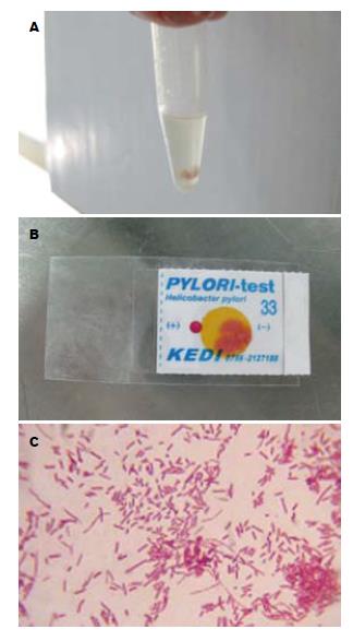
1.2.3 培养并鉴定: 将培养平板置于37℃微需氧环境(装有产气袋的微需氧培养罐)中进行培养, 接种3 d后观察分离效果. 并对细菌进行鉴定: (1)观察细菌形态: 划线接种的H. pylori菌落呈透明针尖样(直径1-2 mm)透明菌落[10], 菌落数可能较少(甚至每块板只有1个), 需仔细对光观察以免遗漏; L棒接种菌量大时, 菌落在培养板表面融合成1层半透明的菌苔. (2)制作湿涂片: 滴1滴生理盐水于干净载玻片上, 用接种环刮取少许固体培养菌苔在生理盐水中涂开. 湿涂片烤干后进行常规Gram染色, 普通显微镜下观察. 典型的H. pylori涂片染色镜检呈Gram阴性海鸥状、S状弯曲菌或短杆菌, 除可见到以上典型形态外还可见到球形体、长丝体等H. pylori形态变异体, 均为Gram阴性染色. 动力好的细菌可观察到典型的钻探样运动. (3)生化鉴定: 尿素酶: 刮取一环细菌置入尿素酶试纸条上, 37℃, 阳性者约1 min后, 试剂应变成红色或紫红色.
1.2.4 传代与增菌: 如培养平板中含有杂菌, 采用分区划线分离单个菌落, 再选取传代培养后的纯化H. pylori菌落或分纯后的单个菌落, 采用密集划线法接种, 进行增菌培养.
1.2.5 保存: 用接种环挑取经革兰染色及生化鉴定已证实的H. pylori良好菌落, 用脱纤维绵羊血低温液态保存, 将固体培养基中细菌用接种环刮取放置在2 mL菌种管(5 mL脱纤维绵羊血)中, 每管置入2-3环细菌. 因H. pylori菌落保存后复苏成功率极低, 为提高复苏率, 故1株菌至少保存3管, 迅速将菌落放置到-80℃冰箱中保存[11].
统计学处理 数据使用SPSS16.0统计软件χ2检验, 以P<0.05为具有显著性差别.
本次研究共采集98例临床标本, 其中37例分离培养结果为阳性, 阳性率为38%. 转运及鉴定过程如图1.
汉族组标本培养80例, 阳性29例, 阳性率36%; 少数民族组标本培养18例, 阳性8例, 阳性率为44%. 少数民族组与汉族组, 2者无显著差异. 其中苗族组培养阳性率与汉族组比较显著升高(P<0.05, 表1).
| 分组 | H. pylori (+) | H. pylori (-) |
| 汉族 | 36 | 64 |
| 苗族 | 100 | 0 |
| 布依族 | 33 | 67 |
| 土家族 | 33 | 67 |
| 白族 | 33 | 67 |
| 侗族 | 0 | 100 |
| 少数民族 | 44 | 56 |
患慢性胃炎伴糜烂及消化性溃疡患者培养阳性率较单纯胃炎、胃癌患者显著升高(均P<0.05), 慢性胃炎伴糜烂及消化性溃疡患者2者间无明显差异(表2).
| 分组 | H. pylori (+) | H. pylori (-) |
| 慢性胃炎 | 0 | 100 |
| 十二指肠炎 | 33 | 67 |
| 慢性胃炎伴糜烂 | 39 | 61 |
| 消化性溃疡 | 46 | 54 |
| 胃癌 | 0 | 100 |
30-60岁年龄段较60-80岁培养阳性率显著增高, 培养阳性率分别为45%、21%(P<0.05), 2者与20-30岁年龄段相比较均无差异性(表3).
| 不同年龄组(岁) | H. pylori (+) | H. pylori (-) |
| 20-30 | 36 | 64 |
| 31-40 | 42 | 58 |
| 41-50 | 57 | 43 |
| 51-60 | 37 | 63 |
| 61-70 | 13 | 87 |
| 71-80 | 33 | 67 |
男性病例培养56例, 阳性27例, 阳性率为48%; 女性病例培养42例, 阳性10例, 阳性率为24%. 男性病例培养阳性率较高(P<0.05).
有胃炎家族史标本培养25例, 阳性9例, 阳性率为36%; 无胃炎家族史标本培养73例, 阳性28例, 阳性率为38%, 两者无显著差异.
H. pylori临床菌株的分离培养是诊断H. pylori感染的"金标准"[12-13], 亦是H. pylori基础及临床研究的一项基本技术. 成功分离培养的临床菌株可用于细菌分型、致病机制研究、构建动物模型、开发免疫疫苗及确定致病因子等基础研究; 亦可用于H. pylori的常规诊断、评价新的诊断方法、体外药敏实验等[14]. 本研究对我省98例上消化系疾病患者的胃黏膜进行H. pylori培养, 取材部位为胃窦, 成功分离38例H. pylori临床菌株, 阳性率为38%. 在我省首次成功分离出H. pylori临床菌株, 对我省H. pylori相关性疾病的基础及临床研究具有重要意义.
为了提高H. pylori的培养阳性率, 我们前期的预实验显示H. pylori培养过程中需注意以下环节: (1)抑制杂菌的生长: 取材过程中, 需要保证取材工具如一次性活检钳、镊子、转送液的无菌; 胃中过路的或寄生的杂菌较多, 在转送液或培养基中需加入选择性的抑菌剂; (2)转送过程中, 常温下标本由内镜室转送到实验室过程不能超过3-4 h, 时间太长或温度过高可能导致细菌死亡; (3)接种标本时, 剪材用的剪刀在火烧灭菌时应冷却后再剪碎标本, 如果温度太高亦会影响培养阳性率; (4)商品化的产气袋与普通的抽气换气法相比较, 可以明显提高培养阳性率, 且方便操作; (5)培养基: 我们加入H. pylori培养添加剂的固体培养基[15-16], 经济实惠; (6)培养时间: 本研究认为H. pylori的最佳培养时限是3 d, 这时菌落的肉眼形态与镜下菌体形态均很典型, 细菌处于对数生长期, 可以进行传代、保存或药物敏感性试验等研究. 约5 d后细菌开始有球形变, 延长培养至7 d时, 由于培养基老化、营养成分缺失等原因, H. pylori形态呈球形变, 且低温保存后难以复苏成功.
在方法学成功培养H. pylori的基础上, 我们进一步探讨了成功分离培养H. pylori临床菌株与入选患者病种、年龄、性别、取材部位、家族史的相关性, 以期后期工作中通过病例选择进一步提高H. pylori临床菌株的分离培养阳性率, 并探讨H. pylori感染与上述因素的相关性.
H. pylori感染与慢性胃炎, 消化性溃疡, 胃癌, 胃黏膜相关淋巴组织淋巴瘤密切相关[17-18]. 本研究亦证实慢性胃炎伴糜烂及消化性溃疡患者培养阳性率较单纯胃炎、胃癌患者显著升高(P<0.05), 胃炎伴糜烂及消化性溃疡患者2者间无差异. 提示H. pylori培养阳性率随胃炎严重程度而升高.
大量研究发现, H. pylori感染存在地区、种族、社会经济差异[19-21]. 本研究结果提示苗族组培养阳性率与汉族组比较显著升高(P<0.05). 贵州地区是少数民族聚居区, 其中以苗族为主要代表, 4名苗族患者H. pylori感染全部呈阳性, 这说明我地区H. pylori感染除受到环境因素影响外, 亦可能与苗族遗传素质或基因型有关, 尚需进一步增加样本例数观察.
H. pylori与性别、年龄的关系, 全世界报告各不相同[22-24]. 我地区以往缺乏H. pylori感染与患者性别相关性的研究. 本研究提示男性H. pylori的阳性率远高于女性的阳性率, 这可能与我地区男性患者的某些生活习惯或遗传素质有关, 尚需进一步研究. 我们的研究亦显示, 30-60岁年龄段H. pylori阳性率较60-80岁者显著升高, 培养阳性率分别为45%、21%(P<0.05), 这可能与老年人免疫功能减退有关. 值得注意的是40岁左右的H. pylori感染较其他年龄段明显升高, 因此H. pylori感染临床菌株监测、疫苗预防或治疗干预可重点在男性40岁左右的患者.
目前认为, H. pylori感染往往有家族聚集性[25-26], 其原因一方面是由于致病性H. pylori可通过粪口或口口途径在家庭内传播[27-29]; 另一方面是宿主或菌种某些基因型有利于H. pylori的定植[30]. 我们的研究提示有无胃炎家族史H. pylori培养阳性率无明显差别, 提示我地区H. pylori感染的主要途径除家庭内传播外, 地区、种族、社会经济差异在我省H. pylori感染中占有重要的地位.
总之, 本研究在我省首次成功分离出H. pylori临床菌株, 苗族、慢性胃炎伴糜烂及消化性溃疡患者、青壮年、男性患者培养阳性率显著提高.
H. pylori是感染率最高的细菌之一. 我国H. pylori感染约50%, 贵州省作为欠发达的多民族聚集地, H. pylori感染率高达60%; 同时, H. pylori耐药越来越严重, 根除难度越来越大, H. pylori的研究急迫且重要. H. pylori的分离培养为深入研究奠定了重要基础.
关玉盘, 教授, 首都医科大学附属北京朝阳医院消化科.
H. pylori临床菌株的分离培养作为H. pylori感染诊断和治疗的基础研究, 受到国内外研究人员广泛关注. 成为根治H. pylori的热点之一.
陆华 et al采用细菌培养法及快速尿素酶法从消化系疾病患者胃黏膜活检标本成功检测出H. pylori, 同时采用哥伦比亚血琼脂培养基培养标本, H. pylori阳性率达50%, 杂菌生长率较低. 王桂月 et al认为尿素酶试验简便实用, 快速灵敏; 细菌培养法分离培养技术条件较高, 需要一定的培养时间, 但是能直接证明H. pylori的存在, 且无假阳性出现, 同时可作为药敏试验, 对不同菌株作遗传因子研究.
本研究采用可靠、简易的划线接种法和细菌培养法, 在贵州省首次成功分离出H. pylori临床菌株, 为贵州省H. pylori的基础及临床研究奠定了基础.
H. pylori的分离培养作为H. pylori感染研究的基础, 在基础研究中可以进一步研究不同临床菌株的特性及基因型; 在临床上可用于探讨耐药问题及指导临床治疗.
划线接种法: 为最常用的分离培养细菌的方法, 通过平板划线后,可使细菌分散生长, 形成单个菌落, 有利于从含有多种细菌的标本中分离出目的菌. 他包含分区划线法和连续划线法.
本文研究是贵州省首次成功分离出H. pylori临床菌株, 为贵州及其他地方关于H. pylori基础及临床研究提供了重要信息和技术参考.
编辑: 李军亮 电编:吴鹏朕
| 1. | Asenjo LM, Gisbert JP. [Prevalence of Helicobacter pylori infection in gastric MALT lymphoma: a systematic review]. Rev Esp Enferm Dig. 2007;99:398-404. [PubMed] |
| 2. | Egi Y, Ito M, Tanaka S, Imagawa S, Takata S, Yoshihara M, Haruma K, Chayama K. Role of Helicobacter pylori infection and chronic inflammation in gastric cancer in the cardia. Jpn J Clin Oncol. 2007;37:365-369. [PubMed] [DOI] |
| 3. | Araújo-Filho I, Brandão-Neto J, Pinheiro LA, Azevedo IM, Freire FH, Medeiros AC. Prevalence of Helicobacter pylori infection in advanced gastric carcinoma. Arq Gastroenterol. 2006;43:288-292. [PubMed] |
| 4. | Gisbert JP. [Helicobacter pylori-related diseases: dyspepsia, ulcer and gastric cancer]. Gastroenterol Hepatol. 2008;31 Suppl 4:18-28. [PubMed] [DOI] |
| 5. | Izzotti A, Durando P, Ansaldi F, Gianiorio F, Pulliero A. Interaction between Helicobacter pylori, diet, and genetic polymorphisms as related to non-cancer diseases. Mutat Res. 2009;667:142-157. [PubMed] [DOI] |
| 6. | Isomoto H, Kawazoe K, Inoue K, Kohno S. Usefulness of the immunological rapid urease test for detection of Helicobacter pylori in patients who are reluctant to undergo endoscopic biopsies. Dig Dis Sci. 2006;51:2302-2305. [PubMed] [DOI] |
| 7. | Yang CS, Cao SY, He XJ, Wang YX, Zhang YL. [Study of correlation between helicobacter pylori infection and hyperammonemia and hepatic encephalopathy in cirrhotic patients]. Zhongguo Weizhongbing Jijiu Yixue. 2007;19:422-424. [PubMed] |
| 8. | Tarkhashvili N, Beriashvili R, Chakvetadze N, Moistsrapishvili M, Chokheli M, Sikharulidze M, Malania L, Abazashvili N, Jhorjholiani E, Chubinidze M. Helicobacter pylori infection in patients undergoing upper endoscopy, Republic of Georgia. Emerg Infect Dis. 2009;15:504-505. [PubMed] [DOI] |
| 10. | Ohno H, Murano A. Serum-free culture of H. pylori intensifies cytotoxicity. World J Gastroenterol. 2007;13:532-537. [PubMed] |
| 11. | Graham DY, Kudo M, Reddy R, Opekun AR. Practical rapid, minimally invasive, reliable nonendoscopic method to obtain Helicobacter pylori for culture. Helicobacter. 2005;10:1-3. [PubMed] [DOI] |
| 12. | Yin Y, He LH, Zhang JZ. Successful isolation of Helicobacter pylori after prolonged incubation from a patient with failed eradication therapy. World J Gastroenterol. 2009;15:1528-1529. [PubMed] [DOI] |
| 13. | Koido S, Odahara S, Mitsunaga M, Aizawa M, Itoh S, Uchiyama K, Komita H, Satoh K, Kuniyasu Y, Yamane T. [Diagnosis of Helicobacter pylori infection: comparison with gold standard]. Rinsho Byori. 2008;56:1007-1013. [PubMed] |
| 14. | Schreiber S, Bücker R, Groll C, Azevedo-Vethacke M, Garten D, Scheid P, Friedrich S, Gatermann S, Josenhans C, Suerbaum S. Rapid loss of motility of Helicobacter pylori in the gastric lumen in vivo. Infect Immun. 2005;73:1584-1589. [PubMed] [DOI] |
| 15. | Vega AE, Cortiñas TI, Mattana CM, Silva HJ, Puig De Centorbi O. Growth of Helicobacter pylori in medium supplemented with cyanobacterial extract. J Clin Microbiol. 2003;41:5384-5388. [PubMed] [DOI] |
| 16. | Walsh EJ, Moran AP. Influence of medium composition on the growth and antigen expression of Helicobacter pylori. J Appl Microbiol. 1997;83:67-75. [PubMed] [DOI] |
| 17. | Backert S, Schwarz T, Miehlke S, Kirsch C, Sommer C, Kwok T, Gerhard M, Goebel UB, Lehn N, Koenig W. Functional analysis of the cag pathogenicity island in Helicobacter pylori isolates from patients with gastritis, peptic ulcer, and gastric cancer. Infect Immun. 2004;72:1043-1056. [PubMed] [DOI] |
| 18. | Machado AM, Figueiredo C, Touati E, Máximo V, Sousa S, Michel V, Carneiro F, Nielsen FC, Seruca R, Rasmussen LJ. Helicobacter pylori infection induces genetic instability of nuclear and mitochondrial DNA in gastric cells. Clin Cancer Res. 2009;15:2995-3002. [PubMed] [DOI] |
| 19. | Goh KL, Parasakthi N. The racial cohort phenomenon: seroepidemiology of Helicobacter pylori infection in a multiracial South-East Asian country. Eur J Gastroenterol Hepatol. 2001;13:177-183. [PubMed] [DOI] |
| 20. | Song MJ, Park DI, Hwang SJ, Kim ER, Kim YH, Jang BI, Lee SH, Ji JS, Shin SJ. [The prevalence of Helicobacter pylori infection in Korean patients with inflammatory bowel disease, a multicenter study]. Korean J Gastroenterol. 2009;53:341-347. [PubMed] [DOI] |
| 21. | Seyda T, Derya C, Füsun A, Meliha K. The relationship of Helicobacter pylori positivity with age, sex, and ABO/Rhesus blood groups in patients with gastrointestinal complaints in Turkey. Helicobacter. 2007;12:244-250. [PubMed] [DOI] |
| 22. | Yücel O, Sayan A, Yildiz M. The factors associated with asymptomatic carriage of Helicobacter pylori in children and their mothers living in three socio-economic settings. Jpn J Infect Dis. 2009;62:120-124. [PubMed] |
| 23. | Kanbay M, Gür G, Arslan H, Yilmaz U, Boyacioglu S. The relationship of ABO blood group, age, gender, smoking, and Helicobacter pylori infection. Dig Dis Sci. 2005;50:1214-1217. [PubMed] [DOI] |
| 25. | Goodman KJ, Correa P. Transmission of Helicobacter pylori among siblings. Lancet. 2000;355:358-362. [PubMed] [DOI] |
| 26. | Wan Y, Xu YY, Xue FB, Fan DM. [Meta-analysis on Helicobacter pylori infection between sex and in family assembles]. Zhonghua Liuxingbingxue Zazhi. 2003;24:54-57. [PubMed] [DOI] |
| 27. | Weyermann M, Rothenbacher D, Brenner H. Acquisition of Helicobacter pylori infection in early childhood: independent contributions of infected mothers, fathers, and siblings. Am J Gastroenterol. 2009;104:182-189. [PubMed] [DOI] |
| 28. | Kurtaran H, Uyar ME, Kasapoglu B, Turkay C, Yilmaz T, Akcay A, Kanbay M. Role of Helicobacter pylori in pathogenesis of upper respiratory system diseases. J Natl Med Assoc. 2008;100:1224-1230. [PubMed] |
| 29. | Salih BA. Helicobacter pylori infection in developing countries: the burden for how long? Saudi J Gastroenterol. 2009;15:201-207. [PubMed] [DOI] |
| 30. | Qiao W, Hu JL, Xiao B, Wu KC, Peng DR, Atherton JC, Xue H. cagA and vacA genotype of Helicobacter pylori associated with gastric diseases in Xi'an area. World J Gastroenterol. 2003;9:1762-1766. [PubMed] |


