修回日期: 2007-12-28
接受日期: 2008-01-06
在线出版日期: 2008-01-18
目的: 研究反流性食管炎(reflux esophagitis, RE)患者幽门螺杆菌(H. pylori)感染率, 感染程度与定植部位及胃炎的活动度, 并对比H. pylori阳性与阴性的RE患者食管炎症的严重程度.
方法: 选取从2007-01/03在我科行胃镜检查证实的RE患者89例, 按年龄, 性别进行配对的方式随机抽取无反流对照组89例, 对比两组H. pylori感染率, 感染部位, 感染程度的差异, 并比较RE组中H. pylori阳性与阴性患者食管炎症的严重程度.
结果: RE组与对照组H. pylori感染率无统计学差异(P = 0.137), 但RE组H. pylori感染程度较对照组轻(P胃体 = 0 .024 , P胃窦 = 0.000), 且RE组胃体炎症严重程度较对照组轻(P = 0.001); H. pylori阳性与阴性的RE患者发生洛杉矶(LA)C级分别占8.3%和18.5%, D级分别占4.2%和12.3%, 但食管炎症的轻重程度无显著性差异(P = 0.353).
结论: H. pylori的感染程度及胃体胃炎的活动性与RE的形成有负相关性.
引文著录: 李渊, 周丽雅, 林三仁, 金珠, 崔荣丽, 何平平. 反流性食管炎与幽门螺杆菌感染的关系. 世界华人消化杂志 2008; 16(2): 171-174
Revised: December 28, 2007
Accepted: January 6, 2008
Published online: January 18, 2008
AIM: To study the prevalence, density and pattern of H. pylori infection and the activity of reflux esophagitis (RE) and to compare the severity of esophagitis in H. pylori-infected and non-infected RE patients.
METHODS: We conducted a prospective, random and case-control study. The condition of H. pylori infection and the activity of gastritis in 89 RE patients and 89 non reflux esophagitis patients were compared. The RE patients were divided into H. pylori-positive group and H. pylori-negative group. The severity of esophagitis was compared between the two groups.
RESULTS: There was no statistic difference in the prevalence of H. pylori infection between the RE and control groups (P = 0.137). However, the density of H. pylori (P = 0.024 in the gastric body and P = 0.000 in the gastric sinus) and the activity of corpus gastritis (P = 0.001) in the RE group were significantly lower than those in the control group. There was no statistic difference in the severity of esophagitis between H. pylori-positive and H. pylori-negative groups (P = 0.353), while the grade LA-C was 8.3% and 18.5% respectively and the grade LA-D was 4.2% and 12.3% respectively. No significant difference was found in the severity of esophagitis.
CONCLUSION: The degree of H. pylori infection and the activity of corpus gastritis are inversely correlated with the formation of RE.
- Citation: Li Y, Zhou LY, Lin SR, Jin Z, Cui RL, He PP. Relationship between H. pylori infection and reflux esophagitis. Shijie Huaren Xiaohua Zazhi 2008; 16(2): 171-174
- URL: https://www.wjgnet.com/1009-3079/full/v16/i2/171.htm
- DOI: https://dx.doi.org/10.11569/wcjd.v16.i2.171
近年来, H. pylori感染及溃疡病等H. pylori相关性疾病的发病率呈下降趋势[1], 而胃食管反流病(gastroesophageal reflux disease, GERD)的发病呈上升趋势. GERD与H. pylori感染的关系尚有争议. 本研究探讨了GERD的主要分型之一反流性食管炎(reflux esophagitis, RE)与H. pylori感染的关系, 揭示在RE发病中H. pylori感染所起的作用.
RE组为选取从2007-01/03在我科行胃镜检查证实的RE患者, 对照组为按年龄, 性别与前组配对的方式随机抽取的欲行胃镜检查, 再经反流性疾病问卷[2](reflux diagnostic questionnaire, RDQ, 又称耐信量表, 取RDQ分值12为诊断临界值, 其敏感性为94.12%, 特异性为50%)判定为无反流的患者, 胃镜检查进一步证实无RE, 食管裂孔疝及Barrett食管; 并注意排除一些可能存在反流的因素如贲门松弛和鳞柱状上皮交界(SCJ)模糊. 两组各89例. 两组的共同排除标准为(1)过去4 wk使用过质子泵抑制剂(PPI), 类固醇激素或非甾体类抗炎药; (2)以前进行过根除H. pylori治疗, 或外科手术; (3)伴消化性溃疡, 胃癌以及幽门梗阻等疾病. 所有患者均在胃体和胃窦各取一块组织进行病理学检查. 2名病理检查医生对胃镜诊断结果不知情.
1.2.1 病理诊断标准: 标本经石蜡切片, HE染色及Warrthin-Starry染色, 组织学诊断根据2000年慢性胃炎研讨会所定的诊断标准. 慢性炎症分级标准: 正常为单核细胞每高倍视野不超过5个, 轻度为慢性炎症细胞较少并局限于黏膜浅层, 不超过黏膜层的1/3, 中度为超过黏膜层的1/3, 但未达到2/3, 重度为占据黏膜全层. 病变程度无、轻、中、重分别定义为0、1、2、3分[3].
1.2.2 H. pylori感染程度分级: 观察胃黏膜黏液层, 表面上皮, 小凹上皮和腺管上皮表面的H. pylori.无: 未见H. pylori; 轻: 偶见或小于标本全长1/3有少数H. pylori; 中: H. pylori分布超过标本全长1/3而未达到2/3或连续的, 薄而稀疏的存在于上皮表面; 重: H. pylori成堆存在, 基本分布于标本全长. 标本全长中扣除肠化区域. 感染程度无、轻、中、重分别定义为0、1、2、3分[3]. RE组食管炎严重程度按洛杉矶标准分级(LA分级)[4-5], 并比较H. pylori阳性与阴性的RE患者的食管炎症的严重程度.
统计学处理 RE组和对照组H. pylori的感染率的比较使用McNemar卡方检验, H. pylori感染程度, 胃炎炎症程度的比较使用秩和检验.
入选RE组共89例患者, 其中LA分级A级33例(占37.1%), B级33例(占37.1%), C级14例(占15.7%), D级9例(占10.1%), 男55例, 女34例, 男:女为1.62:1. 入选对照组的人群为年龄性别与RE组一致的无反流患者.
RE组胃体感染H. pylori阳性者共21例(23.6%), 对照组为32例(36.0%), 两组无显著性差异(χ2 = 2.154, P = 0.118). RE组胃窦感染H. pylori阳性者共20例(22.5%), 对照组为32例(32.6%), 两组无显著性差异(χ2 = 1.641, P = 0.200). 若将胃体和/或胃窦H. pylori阳性定为感染阳性, RE组为24例(27.0%), 对照组为35例(39.3%), 两组无统计学差异(χ2 = 2.326, P = 0.137).
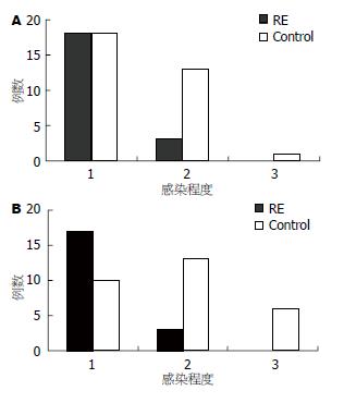
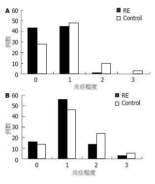
RE组胃体和胃窦H. pylori感染程度较对照组轻(胃体: P = 0.024, Z = -2.251, 胃窦: P = 0.000, Z = -3.545, 图1). RE组的的胃体炎症严重程度较对照组轻(P = 0.001, Z = -3.460, 图2), 而胃窦炎症严重程度与对照组相比差异无显著性(P = 0.076, Z = -1.774, 图2).
RE组中H. pylori阳性者24例, H. pylori阴性者65例, H. pylori阳性及H. pylori阴性患者按LA分级的食管炎症程度比较结果: A级分别占37.5%和36.9%, B级分别占50.0%和32.3%, C级分别占8.3%和18.5%, D级分别占4.2%和12.3%. H. pylori阳性的RE患者食管炎症程度较H. pylori阴性的差异无显著性意义(P = 0.353, Z = -0.0929, 图3).
GERD是一种发病率日渐增加的疾病[6-10], 临床上将他分为NERD(non-erosive reflux disease), RE和Barrett食管, 其中Barrett与食管腺癌的发生密切相关[11]. H. pylori是上消化道的重要致病菌[12-14], 他的感染程度与胃炎的的活动程度呈正相关[15]. 目前流行病学的研究发现, 绝大多数高H. pylori感染国家GERD的患病率低, 而随着这些国家H. pylori感染率的下降, GERD及其并发症的发病率呈上升趋势[8]. 但H. pylori与GERD的关系及其在GERD形成中所起的作用尚不清楚. 本研究通过对RE(GERD的重要分型之一)与无反流对照组胃黏膜H. pylori感染情况及胃炎的活动性进行比较, 来探索GERD和H. pylori的相互关系.
为排除影响H. pylori的各种因素, 并能较为直观地探讨RE和H. pylori的关系, 我们在入组时剔除了使用过PPI类, 抗生素以及消化性溃疡等因素. 我们在内镜检查中发现, GERD患者除了可能有RE, 食管裂孔疝或Barrett典型表现之外, 有时可能仅表现为贲门松弛和SCJ模糊[16], 因此我们使用目前通用的反流性疾病问卷结合内镜检查, 使得无反流组的对照组的选择更可信.
本研究结果显示, RE组与对照组H. pylori感染率相比, 数值上偏低, 但统计学上差异无显著性. 而将胃内H. pylori感染程度进行比较, 则RE组的H. pylori感染程度明显轻于对照组. 若将RE组分成H. pylori感染阳性和感染阴性两组, 比较两组反流的严重程度, 虽统计学上差异无显著性, 但仍能看出H. pylori阳性C级和D级食管炎的病例数较少. 以上结果显示H. pylori与RE的形成呈负相关性. 这与多数研究的结果一致[10,17-19]. 有资料表明RE的H. pylori感染率低于NERD[20], 高度不典型增生的Barrett及食管腺癌的感染率更低[21]. 而且在消化性溃疡及胃炎患者根除H. pylori后, RE的发病率比未根除组高, 从而进一步证实H. pylori对食管有保护性, 而其中H. pylori的作用机制值得探究.
GERD发病机制包括食管抗反流屏障减弱, 食管廓清能力下降, 食管黏膜防御功能异常, 胃酸分泌异常, 胃排空延迟等. 我们的研究发现, RE组的胃体炎症严重程度较对照组轻, 而胃窦炎症严重程度与对照组相比, 差异无显著性. 提示在H. pylori感染后, 当炎症累及胃体时, 胃酸分泌腺体的破坏导致胃酸分泌量的下降[22], 胃内pH值升高[23], 而且升高程度与胃体炎症程度正相关[17]. 另外, 推测H. pylori会产生尿素酶, 分解尿素后产生氨, 氨中和胃酸后使胃内pH值进一步升高, 使食管的酸负荷下降[24], 这会导致GERD发病率下降. 但是尚未发现H. pylori感染影响空腹或餐后下食管扩约肌(lower esophageal sphincter, LES)压力, 也未改变一过性LES松弛的频率, 并且不会改变胃排空[25-27], 因此H. pylori没有破坏抗反流屏障, 他对GERD的形成起的作用相对较弱. 根除H. pylori后也不会增加反流屏障正常者GERD的患病率[18]. 但对反流屏障功能异常的患者, 根除H. pylori后, 胃体炎症减轻, GERD的患病率可能会增加[27-28].
因此, 随着中国H. pylori感染率的下降, GERD及其并发症食管腺癌的发病率有可能呈上升趋势[29], 应引起我们足够的重视, 并应加强研究.
GERD是目前研究热点. 目前的许多流行病学研究显示, H. pylori感染率高的国家GERD发病率较低; 这些国家
H. pylori感染率下降后, GERD发病率有所升高, 而GERD发病率与H. pylori感染率及其他因素, 如生活方式改变的关系如何尚存争议.
伊力亚尔·夏合丁, 教授, 新疆医科大学第一附属医院胸外科
本研究通过对比RE(GERD的亚型之一)和无反流者的病理组织学特点(H. pylori感染和胃炎情况), 显示RE的产生与
H. pylori感染呈一定程度的负相关性.
本文课题设计合理, 数据可靠, 书写规范, 有一定的临床参考价值和学术价值.
编辑: 程剑侠 电编:何基才
| 1. | Zhou LY, Xue Y, Lin SR, Meng LM, Li CF, Yan XE, Gao N, Wang K, Duan ZY. The changes of gastric diseases during the past twenty five years. Zhonghua Nei Ke Za Zhi. 2005;44:431-433. [PubMed] |
| 4. | DeVault KR, Castell DO. Updated guidelines for the diagnosis and treatment of gastroesophageal reflux disease. The Practice Parameters Committee of the American College of Gastroenterology. Am J Gastroenterol. 1999;94:1434-1442. [PubMed] |
| 5. | DeVault KR, Castell DO. Updated guidelines for the diagnosis and treatment of gastroesophageal reflux disease. Am J Gastroenterol. 2005;100:190-200. [PubMed] |
| 6. | Armstrong D. Gastroesophageal reflux disease. Curr Opin Pharmacol. 2005;5:589-595. [PubMed] |
| 7. | Fujimoto K. Review article: prevalence and epidemiology of gastro-oesophageal reflux disease in Japan. Aliment Pharmacol Ther. 2004;20 Suppl 8:5-8. [PubMed] |
| 8. | Ho KY, Cheung TK, Wong BC. Gastroesophageal reflux disease in Asian countries: disorder of nature or nurture? J Gastroenterol Hepatol. 2006;21:1362-1365. [PubMed] |
| 9. | Richter JE. The many manifestations of gastroesophageal reflux disease: presentation, evaluation, and treatment. Gastroenterol Clin North Am. 2007;36:577-599, viii-ix. [PubMed] |
| 10. | Ho KY, Chan YH, Kang JY. Increasing trend of reflux esophagitis and decreasing trend of Helicobacter pylori infection in patients from a multiethnic Asian country. Am J Gastroenterol. 2005;100:1923-1928. [PubMed] |
| 11. | Vakil N, van Zanten SV, Kahrilas P, Dent J, Jones R. The Montreal definition and classification of gastroesophageal reflux disease: a global, evidence-based consensus paper. Z. Gastroenterol. 2007;45:1125-1140. [PubMed] |
| 12. | Correa P, Houghton J. Carcinogenesis of Helicobacter pylori. Gastroenterology. 2007;133:659-672. [PubMed] |
| 13. | Moss SF, Malfertheiner P. Helicobacter and gastric malignancies. Helicobacter. 2007;12 Suppl 1:23-30. [PubMed] |
| 14. | Rokkas T, Simsek I, Ladas S. Helicobacter pylori and non-malignant diseases. Helicobacter. 2007;12 Suppl 1:20-22. [PubMed] |
| 15. | Zhou LY, Shen ZY, Lin SR, Jin Z, Ding SG, Huang XB, Xia ZW, Liu JJ, Guo HL, William C. Changes of gastric mucosa histopathology after Helicobacter pylori eradication. Zhonghua Nei Ke Za Zhi. 2003;42:162-164. [PubMed] |
| 16. | Xue Y, Zhou LY, Lin SR, Huang YH. The application of high-resolution endoscopy in non-erosive reflux disease. Zhonghua Nei Ke Za Zhi. 2006;45:389-392. [PubMed] |
| 17. | Wu JC, Sung JJ, Ng EK, Go MY, Chan WB, Chan FK, Leung WK, Choi CL, Chung SC. Prevalence and distribution of Helicobacter pylori in gastroesophageal reflux disease: a study from the East. Am J Gastroenterol. 1999;94:1790-1794. [PubMed] |
| 18. | Unal S, Karakan T, Dogan I, Cindoruk M, Dumlu S. The influence of Helicobacter pylori infection on the prevalence of endoscopic erosive esophagitis. Helicobacter. 2006;11:556-561. [PubMed] |
| 19. | Zhang J, Chen XL, Wang KM, Guo XD, Zuo AL, Gong J. Relationship of gastric Helicobacter pylori infection to Barrett's esophagus and gastro-esophageal reflux disease in Chinese. World J Gastroenterol. 2004;10:672-675. [PubMed] |
| 20. | Manes G, Mosca S, Laccetti M, Lioniello M, Balzano A. Helicobacter pylori infection, pattern of gastritis, and symptoms in erosive and nonerosive gastroesophageal reflux disease. Scand J Gastroenterol. 1999;34:658-662. [PubMed] |
| 21. | Weston AP, Badr AS, Topalovski M, Cherian R, Dixon A, Hassanein RS. Prospective evaluation of the prevalence of gastric Helicobacter pylori infection in patients with GERD, Barrett's esophagus, Barrett's dysplasia, and Barrett's adenocarcinoma. Am J Gastroenterol. 2000;95:387-394. [PubMed] |
| 22. | Gao BX, Duan LP, Wang K, Xia ZW, Lin SR. The roles of Helicobacter pylori and pattern of gastritis in the pathogenesis of reflux esophagitis. Zhonghua Yi Xue Za Zhi. 2006;86:2674-2678. [PubMed] |
| 23. | Wu JC, Chan FK, Wong SK, Lee YT, Leung WK, Sung JJ. Effect of Helicobacter pylori eradication on oesophageal acid exposure in patients with reflux oesophagitis. Aliment Pharmacol Ther. 2002;16:545-552. [PubMed] |
| 24. | Lee OJ, Lee EJ, Kim HJ. Correlations among gastric juice pH and ammonia, Helicobacter pylori infection and gastric mucosal histology. Korean J Intern Med. 2004;19:205-212. [PubMed] |
| 25. | Tanaka I, Tatsumi Y, Kodama T, Kato K, Fujita S, Mitsufuji S, Kashima K. Effect of Helicobacter pylori eradication on gastroesophageal function. J Gastroenterol Hepatol. 2004;19:251-257. [PubMed] |
| 26. | Graham DY. The changing epidemiology of GERD: geography and Helicobacter pylori. Am J Gastroenterol. 2003;98:1462-1470. [PubMed] |
| 27. | Manes G, Esposito P, Lioniello M, Bove A, Mosca S, Balzano A. Manometric and pH-metric features in gastro-oesophageal reflux disease patients with and without Helicobacter pylori infection. Dig Liver Dis. 2000;32:372-377. [PubMed] |


