修回日期: 2006-05-01
接受日期: 2006-05-08
在线出版日期: 2006-06-28
目的: 探讨螺旋CT对胃癌术前TNM分期的准确性, 指导临床合理地制订治疗方案和进行预后分析.
方法: 术前对45例胃癌患者的腹部SCT资料进行TNM分期, 并与术后病理进行对照研究.
结果: 螺旋CT对胃癌T分期、N分期、M分期和TNM分期的准确率分别为75.6%、73.3%、86.7%和75.6%. 如以平扫CT值≥25 Hu或动脉期CT值≥70 Hu或静脉期CT值≥80 Hu为诊断阳性淋巴结标准, 则阳性淋巴结的敏感性高达98.4%, 特异性为64.0%.
结论: 螺旋CT对胃癌的术前TNM分期可提供较高的准确率.
引文著录: 赵志清, 郑可国, 许达生. 螺旋CT在胃癌术前TNM分期中的应用价值. 世界华人消化杂志 2006; 14(18): 1785-1789
Revised: May 1, 2006
Accepted: May 8, 2006
Published online: June 28, 2006
AIM: To explore the accuracy of spiral computed tomography in the staging of gastric cancer, and to provide clinical guide for the treatment and prognosis analysis.
METHODS: Forty-five patients with gastric cancer received TNM staging by spiral computed tomography before operation. The results were comparatively analyzed with the surgical pathology.
RESULTS: The accuracy of SCT for T stage, N stage and M stage and total TNM staging were 75.6%, 73.3%, 86.7%, and 75.6%, respectively. If the attenuation values of more than or equal to 25 Hu in plain scan, or 70 Hu in arterial phase, or 80 Hu in venous phase were used as a criterion to detect the metastasis-positive lymph nodes, the sensitivity and specificity were 98.4% and 64.0%, respectively.
CONCLUSION: Spiral computed tomography is accurate in the preoperative TNM staging for gastric cancer.
- Citation: Zhao ZQ, Zheng KG, Xu DS. Value of spiral computed tomography in preoperative TNM staging for gastric cancer. Shijie Huaren Xiaohua Zazhi 2006; 14(18): 1785-1789
- URL: https://www.wjgnet.com/1009-3079/full/v14/i18/1785.htm
- DOI: https://dx.doi.org/10.11569/wcjd.v14.i18.1785
胃癌是世界上最常见的恶性肿瘤, 其预后主要与肿瘤的TNM分期、组织学类型和分化程度等有关[1-3]. 能否彻底清除已有转移的淋巴组织是决定患者预后的最主要因素之一, 因此胃癌的术前分期对临床治疗方案的制定、估计预后等有着非常重要的意义[4-7]. 我们探讨螺旋CT(SCT)对胃癌术前TNM分期的准确性, 指导临床合理地制定胃癌的治疗方案和进行预后分析.
2003-2005年胃癌患者45例, 男31例, 女14例. 年龄30-78(平均56)岁. 所有患者术前均经胃镜活检证实, 术后进行病理分期.
CT检查采用的是东芝Xpress/sx螺旋CT机. 全部病例于术前10 d内行螺旋CT检查. CT扫描前患者禁食8-12 h, 检查前口服饮用水800-1000 mL. 扫描层厚0.5 cm, 扫描条件120 kV, 250 mAs, 矩阵512×512, 螺距为1. 先行平扫, 然后行双期增强扫描, 对比剂用碘普胺注射液, Ⅰ浓度为300 g/L, 总量按1.5 mL/kg, 采用EN Vision CT高压注射器, 流率3 mL/s, 经肘静脉注射对比剂后27 s开始动脉期和60 s开始静脉期扫描. 螺旋CT图像术前经两位腹部放射学高年资医师认真观察、测量和术前分期, 并与手术病理进行对照. 胃癌T分期主要参照Lee et al[8]的螺旋CT诊断标准, 胃癌N分期的标准采用国际抗癌联盟(international union control cancer, IUCC)公布的第5版为标准[9]. 淋巴结转移用其长径≥1.0 cm或增强后CT值≥100 Hu为转移阳性的标准[3-10].
统计学处理 测量所得的数据用SPSS 11.0统计学软件进行处理, 各组间的数据以mean±SD表示, 计量资料各组间均数比较用t检验, 计数资料的比较用R×C表资料的χ2检验, 以P<0.01表示差异有统计学意义.
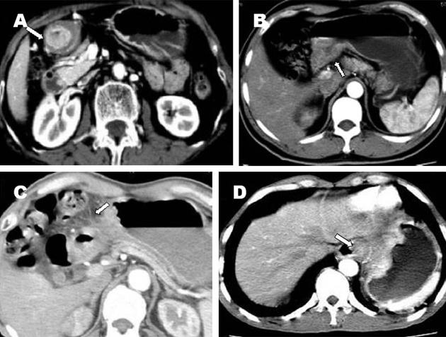
胃癌45例螺旋CT的T分期准确率分别为: T1期50.0%(图1A), T2期66.7%(图1B), T3期83.3%(图1C), T4期75.0%(图1D), 总T分期准确率为75.6%(表1).
| 病理T分期 | 螺旋CT-T分期 | |||
| T1 | T2 | T3 | T4 | |
| T1 | 3 | 2 | 1 | |
| T2 | 2 | 1 | ||
| T3 | 2 | 20 | 2 | |
| T4 | 3 | 9 | ||
胃癌45例螺旋CT的N分期准确率分别为: N0期82.4%, N1期71.4%, N2期71.4%, N3期57.1%. 总的N分期准确率为73.3%(表2).
| 病理N分期 | 螺旋CT-N分期 | |||
| N0 | N1 | N2 | N3 | |
| N0 | 14 | 3 | ||
| N1 | 4 | 10 | ||
| N2 | 2 | 5 | ||
| N3 | 2 | 1 | 4 | |
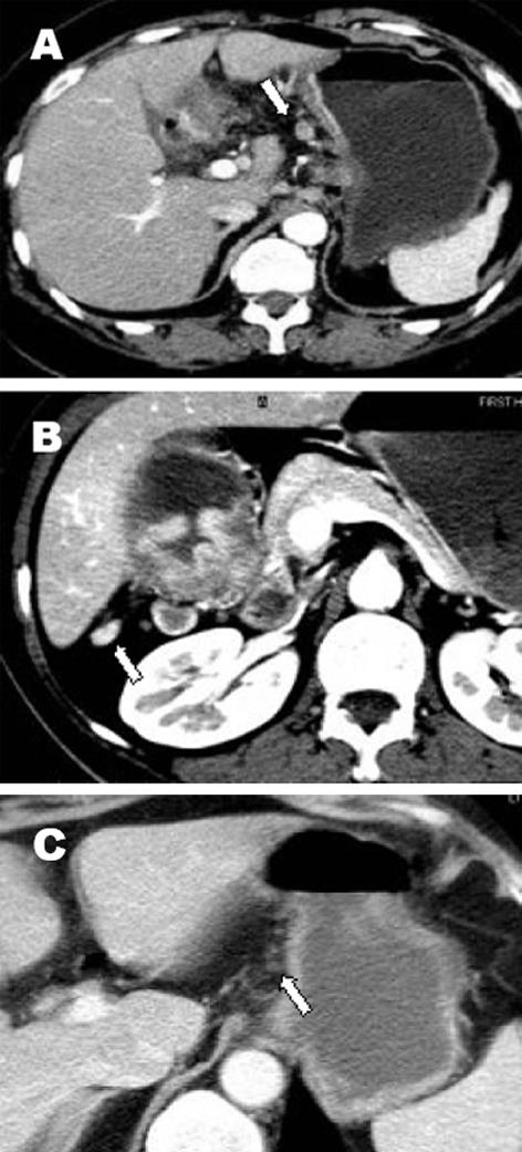
胃癌45例根治性手术者共摘取淋巴结869个. 病理证实淋巴结转移阳性308个, 阳性率为35.4%. 螺旋CT共检出493个淋巴结, 以第3组淋巴结最易被检出(图2A); 术后病理报告为全阳性或全阴性的各组淋巴结共267个, 阳性淋巴结123个(图2A-B), 阴性淋巴结144个(图2C); 其中平扫CT值≥25 Hu或动脉期CT值≥70 Hu或静脉期CT值≥80 Hu的淋巴结共有173个, 其中阳性淋巴结有121个, 阴性淋巴结有52个(表3).
| 淋巴结 | 螺旋CT | 共计 | |
| 阳性淋巴结 | 阴性淋巴结 | ||
| 阳性 | 121 | 2 | 123 |
| 阴性 | 52 | 92 | 144 |
| 共计 | 173 | 94 | 267 |
本组结果显示螺旋CT对T1的准确率较低, 仅50%, 主要是因为相当于黏膜下层的低密度带观察不满意, 误判为肿瘤已侵犯黏膜下层. 螺旋CT对T2的准确率亦较低, 为66.7%, 主要是因为息肉型或隆起型病灶在螺旋CT图像上能见到完整的黏膜下层而误为T1, 而在病理检查时发现病灶已侵犯肌层, 因此确认相当于黏膜下层的低密度带完整性是区分T1和T2的关键, 有时这是很困难的, 因为这层低密度带可能是水肿, 也可能是脂肪沉积引起, 而非真正的黏膜下层[10-13]. 螺旋CT对T3的准确率为83.3%, 相对较高; 误诊Ô因主要有2例胃癌病灶在螺旋CT图像上显示有光滑的边界而误为T2, 1例因胃壁与胰腺之间的脂肪层消失误为T4, 1例因胃壁与肝脏之间的脂肪层消失而误为T4. 螺旋CT对T4的准确率为75.0%, 晚期胃癌常侵犯胰腺、左肝、横结肠及肠系膜等邻近组织、器官, 最常侵犯的脏器是胰腺, 表现为胃胰间的脂肪层消失, 胰腺实质内出现低密度的异常强化灶. 胃癌对肝脏侵犯的CT表现为肿块与肝左叶黏连、分界不清或两者之间的脂肪层消失, 肝左叶实质内出现低密度灶, 在静脉期因正常肝实质明显强化而利于肝脏病灶显示[14-20]. 本组研究提示螺旋CT总的T分期准确率较高(75.6%), 但早期胃癌T分期的准确性偏低, 如何提高早期胃癌的准确分期有待今后进一步研究.
淋巴结转移是胃癌的主要转移方式, 也是影响胃癌术前分期、手术方式选择和预后的一个重要因素, 因而术前影像学判断有无淋巴结转移具有重要的临床意义[21-23]. 螺旋CT常低分胃癌的N分期, 且准确率不高. 主要是螺旋CT估断有无淋巴结转移较困难, 缺乏统一标准, 文献报道主要是依据淋巴结最大直径的大小、短轴与长轴之比、增强时有无强化等为标准. 而各学者的标准又互不相同, 部分学者以淋巴结的最大直径≥0.8-1.5 cm为标准, 但绝大多数以直径≥1.0 cm为标准, 亦有以直径≥0.5 cm为标准者[1-9,13-18]. 以往文献绝大多数都是事先设定淋巴结的直径大于一定的值为转移, 再来评价胃癌淋巴结的转移情况, 众所周知, 标准以上的淋巴结不全都是转移, 而部分转移的淋巴结的直径并不一定增大. 我们以平扫和增强后淋巴结的CT值为依据来判断淋巴结是否转移, 可见本组术后病理诊断为全阳组或全阴组的淋巴结共267个, 如以平扫CT值≥25 Hu或动脉期CT值≥70 Hu或静脉期CT值≥80 Hu为诊断阳性淋巴结标准, 则阳性淋巴结的敏感性高达98.4%, 特异性约为64.0%; 阳性淋巴结的敏感性和特异性均较理想, 值得推荐.
胃癌的M分期是指胃癌远处脏器的转移情况, 主要有腹腔内种植转移、血行转移. 当胃癌种植到卵巢时, 叫Krukenberg瘤, 常见于胃印戒细胞癌. 腹膜转移CT表现为网膜、系膜脂肪密度增高、模糊、腹壁结节、网膜饼、污垢样腹膜及腹水等. 胃癌血行转移时, 最常见的是转移至肝, 其次为肺、肾上腺、肾、骨和脑[24-25]. 本组45例胃癌中M1期11例, 其中有4例手术发现有盆腔转移, 因仅作上腹部CT扫描, 而误为M0期, 有2例腹膜转移但无腹水患者, 在CT图像上因结节细小未发现, 而误为M0期. 胃癌总的TNM分期的准确率取决于各分期的准确率, 尤其是T分期与N分期的准确率. 本组结果显示, 螺旋CT具有较高的TNM分期准确率(75.6%), 但对早期胃癌(Ⅰ, Ⅱ期胃癌)的分期准确率较低, 主要与螺旋CT对早期胃癌的T分期准确率较低有关. 从目前发表的文献来看, 螺旋CT对胃癌总的TNM分期的准确率较EUS, 普通CT, MRI的准确率高[26-27], 且为无创性检查, 因此, 螺旋CT是胃癌治疗前进行准确分期的首选检查方法.
总之, 胃癌的术前螺旋CT检查具有较高的TNM分期准确率, 目前已成为胃癌术前分期的一项常规检查. 应用平扫CT值≥25 Hu或动脉期CT值≥70 Hu或静脉期CT值≥80 Hu为诊断阳性淋巴结标准, 可提高对阳性淋巴结的判断. 但对小淋巴结是否转移尚有一定的难度, 有待今后进一步研究.
胃癌是世界上最常见的恶性肿瘤, 能否彻底清除已有转移的淋巴组织是决定患者预后的最主要因素之一, 因此胃癌的术前分期对临床治疗方案的制定、估计预后等有着非常重要的意义. 本文旨在探讨SCT对胃癌术前TNM分期的准确性, 指导临床合理地制定胃癌的治疗方案和进行预后分析.
尽管有关螺旋CT在胃癌TNM分期中的研究国内外已见报道, 但本文研究结果对指导胃癌治疗方案及预后评估仍有一定意义. 文章逻辑性较强, 数据客观, 影像资料真实, 有一定学术价值.
电编: 张敏 编辑:张海宁
| 1. | Sohn KM, Lee JM, Lee SY, Ahn BY, Park SM, Kim KM. Comparing MR imaging and CT in the staging of gastric carcinoma. AJR Am J Roentgenol. 2000;174:1551-1557. [PubMed] [DOI] |
| 2. | Gossios K, Tsianos E, Prassopoulos P, Papakonstan-tinou O, Tsimoyiannis E, Gourtsoyiannis N. Usefulness of the non-distension of the stomach in the evaluation of perigastric invasion in advanced gastric cancer by CT. Eur J Radiol. 1998;29:61-70. [PubMed] [DOI] |
| 3. | Motohara T, Semelka RC. MRI in staging of gastric cancer. Abdom Imaging. 2002;27:376-383. [PubMed] [DOI] |
| 4. | Tunaci M. Carcinoma of stomach and duodenum: radiologic diagnosis and staging. Eur J Radiol. 2002;42:181-192. [PubMed] [DOI] |
| 5. | Lim JS, Yun MJ, Kim MJ, Hyung WJ, Park MS, Choi JY, Kim TS, Lee JD, Noh SH, Kim KW. CT and PET in stomach cancer: preoperative staging and monitoring of response to therapy. Radiographics. 2006;26:143-156. [PubMed] [DOI] |
| 6. | Kim AY, Kim HJ, Ha HK. Gastric cancer by multidetector row CT: preoperative staging. Abdom Imaging. 2005;30:465-472. [PubMed] [DOI] |
| 7. | Tschmelitsch J, Weiser MR, Karpeh MS. Modern staging in gastric cancer. Surg Oncol. 2000;9:23-30. [PubMed] [DOI] |
| 8. | Lee DH, Seo TS, Ko YT. Spiral CT of the gastric carcinoma: staging and enhancement pattern. Clin Imaging. 2001;25:32-37. [PubMed] [DOI] |
| 9. | Yoo CH, Noh SH, Kim YI, Min JS. Comparison of prognostic significance of nodal staging between old (4th edition) and new (5th edition) UICC TNM classification for gastric carcinoma.International Union Against Cancer. World J Surg. 1999;23:492-497; discussion 497-498. [PubMed] |
| 10. | Nishimata H, Maruyama M, Shimaoka S, Nishimata Y, Ohi H, Niihara T, Nioh T, Matsuda A, Tashiro K, Torimaru H. Early gastric carcinomas in the cardiac region: diagnosis with double-contrast x-ray studies. Abdom Imaging. 2003;28:486-491. [PubMed] [DOI] |
| 11. | Kim HJ, Kim AY, Oh ST, Kim JS, Kim KW, Kim PN, Lee MG, Ha HK. Gastric cancer staging at multi-detector row CT gastrography: comparison of transverse and volumetric CT scanning. Radiology. 2005;236:879-885. [PubMed] [DOI] |
| 12. | Gamon Giner R, Escrig Sos J, Salvador Sanchis JL, Ruiz del Castillo J, Garcia Vila JH, Marcote Valdivieso E. Helical CT evaluation in the preopera-tive staging of gastric adenocarcinoma. Rev Esp Enferm Dig. 2002;94:593-600. [PubMed] |
| 13. | Stabile Ianora AA, Pedote P, Scardapane A, Memeo M, Rotondo A, Angelelli G. Preoperative staging of gastric carcinoma with multidetector spiral CT. Radiol Med (Torino). 2003;106:467-480. [PubMed] |
| 14. | Shimizu K, Ito K, Matsunaga N, Shimizu A, Kawakami Y. Diagnosis of gastric cancer with MDCT using the water-filling method and multipla-nar reconstruction: CT-histologic correlation. AJR Am J Roentgenol. 2005;185:1152-1158. [PubMed] [DOI] |
| 15. | Matsushita M, Oi H, Murakami T, Takata N, Kim T, Kishimoto H, Nakamura H, Okamoto S, Okamura J. Extraserosal invasion in advanced gastric cancer: evaluation with MR imaging. Radiology. 1994;192:87-91. [PubMed] [DOI] |
| 16. | D'Elia F, Zingarelli A, Palli D, Grani M. Hydro-dynamic CT preoperative staging of gastric cancer: correlation with pathological findings. A prospective study of 107 cases. Eur Radiol. 2000;10:1877-1885. [PubMed] [DOI] |
| 17. | Kumano S, Murakami T, Kim T, Hori M, Iannaccone R, Nakata S, Onishi H, Osuga K, Tomoda K, Catalano C. T staging of gastric cancer: role of multi-detector row CT. Radiology. 2005;237:961-966. [PubMed] [DOI] |
| 18. | Wei WZ, Yu JP, Li J, Liu CS, Zheng XH. Evaluation of contrast-enhanced helical hydro-CT in staging gastric cancer. World J Gastroenterol. 2005;11:4592-4595. [PubMed] [DOI] |
| 19. | Kang BC, Kim JH, Kim KW, Lee DY, Baek SY, Lee SW, Jung WH. Value of the dynamic and delayed MR sequence with Gd-DTPA in the T-staging of stomach cancer: correlation with the histopathology. Abdom Imaging. 2000;25:14-24. [PubMed] [DOI] |
| 20. | Yun M, Lim JS, Noh SH, Hyung WJ, Cheong JH, Bong JK, Cho A, Lee JD. Lymph node staging of gastric cancer using (18)F-FDG PET: a comparison study with CT. J Nucl Med. 2005;46:1582-1588. [PubMed] |
| 21. | Mani NB, Suri S, Gupta S, Wig JD. Two-phase dynamic contrast-enhanced computed tomography with water-filling method for staging of gastric carcinoma. Clin Imaging. 2001;25:38-43. [PubMed] [DOI] |
| 22. | Rossi M, Broglia L, Maccioni F, Bezzi M, Laghi A, Graziano P, Mingazzini PL, Rossi P. Hydro-CT in patients with gastric cancer: preoperative radiologic staging. Eur Radiol. 1997;7:659-664. [PubMed] [DOI] |
| 23. | Lakadamyali H, Oto A, Akmangit I, Abbasoglu O, Sivri B, Akhan O, Besim A. The role of spiral CT in the preoperative evaluation of malignant gastric neoplasms. Tani Girisim Radyol. 2003;9:345-353. [PubMed] |
| 24. | Cereceda Perez CN, Urbasos Pascual MI, Romero Castellanos C, Carreira Gomez C, Pinto Varela JM. Helical CT of the stomach: differentiation between benign and malignant pathologies, together with the staging of gastric carcinoma. Rev Esp Enferm Dig. 2002;94:601-612. [PubMed] |
| 25. | Blackshaw GR, Stephens MR, Lewis WG, Boyce J, Barry JD, Edwards P, Allison MC, Thomas GV. Progressive CT system technology and experience improve the perceived preoperative stage of gastric cancer. Gastric Cancer. 2005;8:29-34. [PubMed] [DOI] |
| 26. | Habermann CR, Weiss F, Riecken R, Honarpisheh H, Bohnacker S, Staedtler C, Dieckmann C, Schoder V, Adam G. Preoperative staging of gastric adenocarcinoma: comparison of helical CT and endoscopic US. Radiology. 2004;230:465-471. [PubMed] [DOI] |
| 27. | Chen F, Ni YC, Zheng KE, Ju SH, Sun J, Ou XL, Xu MH, Zhang H, Marchal G. Spiral CT in gastric carcinoma: comparison with barium study, fiberoptic gastroscopy and histopathology. World J Gastroenterol. 2003;9:1404-1408. [PubMed] [DOI] |


