修回日期: 2005-08-20
接受日期: 2005-08-26
在线出版日期: 2005-10-15
目的: 评价生理盐水冲洗技术对于改善多层螺旋CT三维门脉造影MIP图像质量的临床应用价值.
方法: 58例疑有肝脏转移瘤的患者, 随机分为A, B组. A组给与2.0 mL/kg对比剂及30 mL盐水冲洗; B组仅给与等量的对比剂. 对比剂注射时间均为33 s, 盐水的注射速度为3 mL/s. 扫描延迟45 s, 原始数据采用1 mm间隔重建. 测量原始横断图像中肝右叶实质、门脉右干、门脉主干以及腹主动脉增强后的CT值采用4分评定法评估最大密度投影法(MIP)图像质量.
结果: A组的门脉主干、门脉右干的CT值以及门脉右支与肝右叶CT值的差异均较B组高, 两组的差值分别为25.0、19.7、17.6 Hu(P = 0.006, 0.047 及 0.042); A组MIP图像质量为优或者良的比例为60.7% (17/28), 而B组为33.3% (10/30), 两组的3D-CTP MIP图像质量平均评分差值为0.59(P = 0.040), 差异均具有统计学意义.
结论: 生理盐水冲洗技术可以提高门脉对比强化程度、改善MIP图像的质量.
引文著录: 肖亮, 徐克. 生理盐水冲洗提高多层CT门脉造影图像质量的临床研究. 世界华人消化杂志 2005; 13(19): 2343-2348
Revised: August 20, 2005
Accepted: August 26, 2005
Published online: October 15, 2005
AIM: To evaluate the clinical application of normal saline flush technique in improving the three-dimensional image quality of multi-detector helical CT-photography obtained by maximum intensity projection (MIP).
METHODS: Fifty-eight patients were randomly divided into two groups. Patients in the two groups were both injected with 2.0 mL/kg contrast material (300 gI/L), and patients in group A were also treated with 30 mL saline flush (3 mL/s). The injection of the contrast material lasted 33 s in both groups. The scanning was performed 45 s after injection. The scanning started at the level of the diaphragm and covered the entire liver and spleen. The acquired raw data were reconstructed at an interval of 1 mm. The CT values of the right hepatic lobe (RHL), main portal vein (MPV), right portal vein (RPV), and abdominal aorta were assessed. MIP images of 3D-CTP were visually graded by the four point-scoring system.
RESULTS: The mean CT attenuation values of MPV, RPV, and RPV-RHL were higher in group A. The differences between the two groups were 25.0, 19.7, and 17.6 Hu (P = 0.006, 0.047, and 0.042, respectively). The rates of the excellent or good MIP images were 60.7% (17/28) in group A, and 33.3% (10/30) in group B. The mean score of the differences was 0.59, which was significant between the two groups (P = 0.040).
CONCLUSION: The saline flush technique can increase the CT attenuation value of portal vein as well as improve the quality of its MIP images.
- Citation: Xiao L, Xu K. Clinical application of normal saline flush in multi-detector CT photography on portal vein. Shijie Huaren Xiaohua Zazhi 2005; 13(19): 2343-2348
- URL: https://www.wjgnet.com/1009-3079/full/v13/i19/2343.htm
- DOI: https://dx.doi.org/10.11569/wcjd.v13.i19.2343
随着现代多层螺旋CT技术的出现和非离子型对比剂的普遍应用, 三维CT血管造影(3D-CTA)已经成为了一种了解各处血管结构的重要的非创性临床检查手段[1-8]. 与3D-CT动脉造影相比, 3D-CT门脉造影(3D-CTP)由于相对较低的对比增强幅度, 门静脉系统的成像较困难. 因此, 如何提高3D-CTP图像质量一直是放射科医生和临床医生关注的热点. 3D-CTP的图像质量好坏取决于多方面因素[9-18,21-23]. 目前的研究内容主要包括: 重建方法、对比剂应用以及扫描时相的选择. Nakayama et al[21]报道最大密度投影法(MIP)是三种重建方法中门脉及其分支显示最好的一种方法. 关于对比剂的浓度、剂量、注入速度以及扫描时相对3D-CTP图像质量的影响现已有多篇文章报道[22,23], 而注药方式对于3D-CTP图像质量的影响仍不清楚. 当采用团注加生理盐水冲洗的注药方式时, 盐水推动末尾部分对比剂, 从而减少了对比剂的拖尾及稀释, 同时还避免了对比剂滞留于上肢静脉及其造成的胸廓入口部的条带状伪影. 目前, 国外已有一些关于生理盐水冲洗技术减低胸、腹部增强CT对比剂使用剂量的研究报道[19,20,24-30], 国内仍未见类似报道. 我们的目的在于评估生理盐水冲洗技术对于改善3D-CTP图像质量的临床应用价值.
收集2003-05/2004-01中国医科大学附属第一医院58例因怀疑有肝内转移瘤而行腹部增强CT扫描的患者, 男38例, 女21例, 平均年龄为60.3(34-80)岁, 平均体重为57.6 kg. 随机分为A、B两组, 两组间年龄及体重无显著性差异(P = 0.51, 0.839). 一般状态差、严重肝硬化、门静脉血栓形成、肝动脉或门静脉受肿瘤侵袭或压迫、心功能不全的患者均排除于本研究之外. 采用的方法符合我院的临床常规标准. 所有患者均于检查前签署了书面的同意书, 本研究得到了学术研究委员会的批准.
1.2.1 注药及扫描程序: 所有检查均采用18 G静脉套管针穿刺肘前静脉或近端前臂静脉, 于注射对比剂之前, 在扫描体位下, 手推少量生理盐水确保静脉通路的通畅性. 生理盐水及对比剂的注入采用双头的高压注射器, 采用非离子型对比剂欧乃派克, 两组注射药物的程序见表1.
| n | 年龄(岁) | 体重(kg) | 对比剂 | 生理盐水 | ||||
| 剂量 | 注射速度 | 注射时间 | 剂量 | 注射速度 | ||||
| A组 | 28 | 59.32 ± 12.5 | 57.3±9.3 | 2.0 mL/kg | 3.47±0.57 mL/s | 33 s | 30 mL | 3.0 mL/s |
| B组 | 30 | 61.13 ± 7.95 | 57.8±11.0 | 2.0 mL/kg | 3.50±0.67 mL/s | 33 s | 0 | 0 |
所有CT扫描均使用GE LIGHT SPEED 16层螺旋CT机. 扫描条件为0.5 s/螺旋, 扫描层厚0.75 mm, Pitch 12, 管电压120 kV, 管电流150 Eff.mAs. 于注射对比剂45 s后开始扫描, 扫描范围上起膈顶, 向下包括肝、脾的下极, 扫描时间为8-10 s. 原始数据采用1 mm间隔重建并传输到工作站. 采用MIP显示三维CT门脉造影图像.
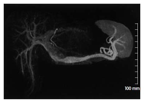
1.2.2 测量CT值及图像评价: 分别采用感兴趣区(ROI)测量原始横断图像中肝右叶实质、门脉右干、门脉主干以及腹主动脉增强后的CT值. 测量者不知道分组的情况. 肝右叶实质的ROI选择在无病灶区域, 内部密度均匀、无可见的血管结构, 尽量保持ROI面积一致, 约100 mm2大小; 血管的ROI应尽量包括全部血管断面. MIP图像由两位不清楚分组情况的观察者按照如下标准进行评估打分: 优(4分)-门脉主干至三级以上分支被清晰显示; 良(3分)-门脉主干至二级分支被清晰显示, 三级分支未被清晰显示; 可(2分)-仅门脉主干或一级分支被清晰显示; 差(1分)-门脉主干未被清晰显示.
统计学处理 两组间各均值的差异采用t检验, 双尾法评价P值, P<0.05被认为有统计学差异.
所有检查过程均未出现药物过敏及对比剂外渗现象. 门脉主干、腹主动脉、门脉右干、肝右叶的CT值以及门脉右支与肝右叶CT值的差异的均数及其标准差见表2.
| 门脉主干 | 腹主动脉 | 门脉右支 | 肝右叶 | 门脉右支与肝右叶之差 | |
| A组 | 231.9 ± 40.2 | 273.0 ± 61.9 | 210.9 ± 34.0 | 98.5 ± 18.7 | 112.5 ± 35.5 |
| B组 | 206.9 ± 24.8 | 252.3 ± 61.0 | 191.2 ± 27.6 | 96.3 ± 14.3 | 94.9 ± 28.5 |
| P值 | 0.006 | 0.205 | 0.047 | 0.610 | 0.042 |
对表2的数据进行分析可以得出两项结果. 第一, 采用生理盐水冲洗技术(A组)所得到的门脉主干、门脉右干的CT值以及门脉右支与肝右叶CT值的差异均较单独使用对比剂(B组)高, 差值分别为25.0、19.7、17.6 Hu, P值分别为0.006、0.047、0.042, 差异具有统计学意义. 第二, 腹主动脉以及肝右叶的CT值两组间的差值分别为20.7和2.2 Hu, P值分别为0.205和0.610, 差异不具有统计学意义.
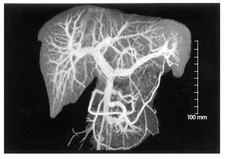
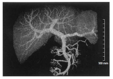
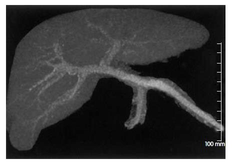
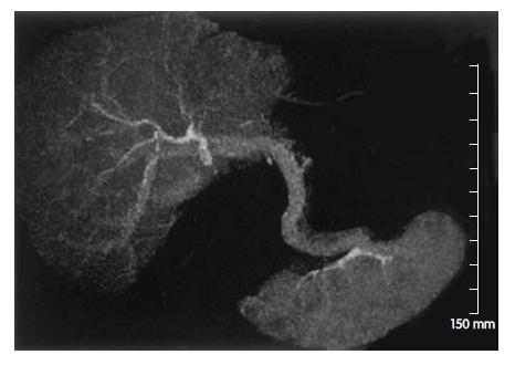
对于3D-CTP MIP图像质量的定性评价及评分结果见表3. 3D-CTP MIP图像质量优、良、可、差的典型图像(图1-5). A组中, MIP图像质量为优或者良的比例为60.7% (17/28); 可或者差的比例为39.3% (11/28). B组中, MIP图像质量为优或者良的比例为33.3% (10/30); 可或者差的比例为66.7% (20/30). 两组平均评分差值为0.59, P值为0.040, 差异具有统计学意义, 3D-CTP图像质量与相关因素的关系见表4.
| 优 | 良 | 可 | 差 | |
| n | 15 | 12 | 19 | 12 |
| 平均年龄 (岁) | 55.67± 14.9 | 61.75 ± 9.8 | 63.11± 6.7 | 60.00± 7.7 |
| 平均体质量 (kg) | 53.00± 10.4 | 55.75 ± 6.3 | 58.74± 9.6 | 63.25±11.7 |
| 门脉主干平均CT值 (Hu) | 246.06± 29.4 | 235.91± 25.6 | 208.57± 28.6 | 184.28± 22.6 |
| 腹主动脉平均CT值 (Hu) | 230.42± 46.6 | 254.44± 58.5 | 275.55± 64.3 | 288.83±65.7 |
| 门脉右支平均CT值 (Hu) | 237.40±23.7 | 217.10±26.4 | 183.30±32.9 | 166.10± 27.4 |
| 肝右叶平均CT值 (Hu) | 106.30±16.5 | 101.30± 15.6 | 93.40±14.4 | 88.50±15.4 |
| 门脉右支与肝右叶CT值之差(Hu) | 131.07± 27.8 | 115.79±18.5 | 89.96±27.5 | 77.58± 28.9 |
3D-CTP的图像质量好坏取决于多方面因素: (1)有无呼吸运动伪影; (2)重建方法的差异; (3)门脉与周围组织之间的X线衰减系数的差异程度. 多层螺旋CT的扫描时间短, 在不足10 s的时间内完成3D-CTP的扫描数据采集过程, 因而呼吸运动伪影通常可以被避免. 3D-CTP的重建方法有3种: MIP、VR、SSD. Nakayama et al[21]报道MIP是3种重建方法中门脉及其分支显示最好的一种方法, 可以比经动脉门脉造影发现更多的门脉侧枝. 影响门脉与其周围组织之间密度差异程度的条件分为内在因素和外加条件两类. 前者主要是门脉的血流状态(血流速度、血流量、流入压力、流出阻力及侧枝循环开放情况), 这与受试者的年龄、体质量等自然情况以及门脉压力有无病理性升高密切相关; 后者包括对比剂的浓度、剂量、注入速度、注药方式以及扫描时相等. Megibow et al[31]采用不同剂量Iopromide(300 gI/L)进行的肝脏增强CT研究, 在使用1.5 mL/kg的剂量下, 肝实质CT值增加量超过30 Hu, 达到良好的强化效果. 我们的研究中采用2.0 mL/kg的剂量, 因为目的是门脉血管结构的显示, 所以需要更多的对比剂. 研究结果表明, 2.0 mL/kg的剂量可以使大多数受试者的门脉结构得到清晰地显示. Yamamoto et al[23]采用300 gI/L的非离子型对比剂100 mL, 对肝硬化患者行3D-CTP检查, 分别延迟30、60、90 s采集原始数据, 得到的结论是延迟60 s扫描时门脉结构显示最清楚. 本研究中, 采用延迟45 s扫描, 基于两方面的原因: (1)受试者均无肝硬化及门脉高压病史, 门脉血流状态好于肝硬化患者; (2)扫描过程处于门脉最高峰之前, 通过分析数据, 可以粗略地判断不同受试者门脉强化达峰时间的早晚.
增强CT的注药方式分为外周静脉单纯团注及团注加生理盐水冲洗两种. 单纯团注时, 末尾部分的对比剂进入体循环缓慢或滞留于外周静脉. 在3D-CTP扫描时, 这部分对比剂处于外周静脉、中央静脉、心脏以及肺部血管床等无效区域内, 不会增加扫描区域血管与周围组织的密度差异. 而在团注加生理盐水冲洗时, 盐水推动末尾部分对比剂, 从而减少了对比剂的拖尾及稀释, 使主动脉时间密度曲线中高峰平台期更长、斜坡走行更陡峭[29]. 我们的研究结果表明, 在对比剂浓度、剂量、注射时间及速度一致以及扫描条件、时间固定不变的情况下, 生理盐水冲洗技术可以提高门脉主干CT值约25.7 Hu, 提高门脉右支与肝右叶实质强化后的密度差异约17.5 Hu, 并显著提高3D-CTP MIP图像的质量. 本研究中, 扫描起始时间与对比剂注射起始时间间隔为45 s, 全部扫描过程约为10 s, 对比剂注射时间为33 s, 根据Bae et al[22]的研究报告, 本研究的扫描过程均处于主动脉强化最高峰与门脉强化最高峰之间. 此阶段的时间密度曲线具有如下特点: (1)主动脉CT值随时间推移而下降, 门脉CT值随时间推移而上升; (2)主动脉与门脉CT值之差随时间推移而下降, 此差值越大, 距门脉强化最高峰越远. 由于我们采用统一的扫描时间间隔, 并没有进行同层动态扫描, 故无法得到时间密度曲线, 不能准确地判断受试者的体质量、年龄与门脉强化达到最高峰的时间的关系, 但通过对实验数据的分析, 可以定性地推断出它们之间的关系. 将全部数据按MIP图像质量的好坏分为优、良、可、差4组进行比较发现MIP图像质量越好, 门脉主干CT值以及门脉右支与肝右叶实质密度差值越大, 而主动脉CT值越低, 受试者平均体重及年龄也越小. 结合上述时间密度曲线的特点可以推断出MIP图像质量越好, 受试者平均体质量及年龄越小, 扫描起始时间距门脉达峰时间越近(门脉达峰时间越早).
本研究中, 所有检查均由同一台16层螺旋CT机完成, 扫描条件相同, 避免了Levi et al[32]报道的使用不同CT扫描机可能造成的测量误差. 他们报道从不同CT扫描机(甚至制造商和型号相同)得到的CT绝对值具有显著性差异.
截止目前, 据我们了解, 关于生理盐水冲洗技术用于胸、腹部增强CT的研究报道并不多见. Hopper et al[25]报道在胸部增强CT中, 采用75 mL对比剂加上生理盐水冲洗与125 mL对比剂无生理盐水冲洗的效果相同. 他们采用的是单筒注射器, 生理盐水被装载于对比剂的上方. Haage et al[24]采用双筒注射器生理盐水冲洗技术, 在肺动脉主干及升主动脉强化效果不受影响的情况下, 将370 gI/L的高浓度对比剂的剂量由75 mL减至60 mL, 同时减低了上腔静脉的伪影发生率. Irie et al[29]报道当进行胸部CT检查时, 采用生理盐水冲洗技术可以延长主动脉高峰平台期持续时间6 s, 同时节省对比剂12 mL. 在最新的一个研究中, Schoellnast et al[27]报道采用300 gI/L非离子型对比剂100 mL及生理盐水冲洗技术使肝实质、门脉、主动脉的CT值比未给予生理盐水冲洗时分别升高9、10、10 Hu. 与之相比, 我们的研究和他们的研究均采用双筒注射器, 组间对比剂剂量一致, 均得出生理盐水冲洗技术使门脉、主动脉CT值升高的结论. 他们采用同一组患者、不同时期的两次增强CT扫描, 延迟时间为70 s, 而我们采用随机分组的两组患者进行研究, 延迟时间为45 s, 并对3D-CTP图像质量进行评估.
本研究的局限之一是对比剂注射起始时间与扫描起始时间间隔固定不变. 因为血液循环速度存在个体差异, 主动脉、门脉对比剂强化的达峰时间因人而异. 本研究中, 部分病例在扫描时门脉强化程度没有达到最大值, 影响了3D-CTP的图像质量. 另外, 在部分病例中, 由于脾静脉及肠系膜上静脉强化程度的差异, 使两者血液汇合时, 出现门脉主干内密度不均匀, 而使MIP图像中门脉主干边缘模糊; 而门脉分支密度均匀, 显影清晰(图5). 对于这种情况我们均将其评为差. 这是本研究中图像差的比例偏大的主要原因. 第二, 我们仅对3D-CTP的MIP图像进行评估. 来源于同一原始数据、采用不同的重建方法得到的3D-CTP图像质量有所不同. 因此, 本研究中所观察到的生理盐水冲洗技术对3D-CTP的MIP图像质量的提高并不意味能对3D-CTP的容积重建(VR)图像产生同样的效果. 关于VR图像的质量改善需要进行进一步的研究.
总之, 生理盐水冲洗技术可以提高门脉对比强化程度, 加大门脉与周围组织的密度差异, 改善3D-CTP MIP图像的质.
电编: 张敏 编辑:潘伯荣 审读:张海宁
| 1. | Schoenhagen P, Halliburton SS, Stillman AE, Kuzmiak SA, Nissen SE, Tuzcu EM, White RD. Noninvasive imaging of coronary arteries: current and future role of multi-detector row CT. Radiology. 2004;232:7-17. [PubMed] [DOI] |
| 2. | Kanne JP, Lalani TA. Role of computed tomography and magnetic resonance imaging for deep venous thrombosis and pulmonary embolism. Circulation. 2004;109:I15-I21. [PubMed] [DOI] |
| 3. | Catalano C, Napoli A, Fraioli F, Venditti F, Votta V, Passariello R. Multidetector-row CT angiography of the infrarenal aortic and lower extremities arterial disease. Eur Radiol. 2003;13 Suppl 5:M88-M93. [PubMed] [DOI] |
| 4. | Schoepf UJ, Costello P. CT angiography for diagnosis of pulmonary embolism: state of the art. Radiology. 2004;230:329-337. [PubMed] [DOI] |
| 5. | Lawler LP, Fishman EK. Multidetector row computed tomography of the aorta and peripheral arteries. Cardiol Clin. 2003;21:607-629. [PubMed] [DOI] |
| 6. | Kang HK, Jeong YY, Choi JH, Choi S, Chung TW, Seo JJ, Kim JK, Yoon W, Park JG. Three-dimensional multi-detector row CT portal venography in the evaluation of portosystemic collateral vessels in liver cirrhosis. Radiographics. 2002;22:1053-1061. [PubMed] [DOI] |
| 7. | Phillips CD, Bubash LA. CT angiography and MR angiography in the evaluation of extracranial carotid vascular disease. Radiol Clin North Am. 2002;40:783-798. [PubMed] [DOI] |
| 8. | Urban BA, Ratner LE, Fishman EK. Three-dimensional volume-rendered CT angiography of the renal arteries and veins: normal anatomy, variants, and clinical applications. Radiographics. 2001;21:373-86; questionnaire 549-55. [PubMed] [DOI] |
| 9. | Bae KT, Tran HQ, Heiken JP. Uniform vascular contrast enhancement and reduced contrast medium volume achieved by using exponentially decelerated contrast material injection method. Radiology. 2004;231:732-736. [PubMed] [DOI] |
| 10. | Maher MM, Kalra MK, Sahani DV, Perumpillichira JJ, Rizzo S, Saini S, Mueller PR. Techniques, clinical applications and limitations of 3D reconstruction in CT of the abdomen. Korean J Radiol. 2004;5:55-67. [PubMed] [DOI] |
| 11. | Martí-Bonmatí L, Tobarra E, Manjón JV, Robles M, Arana E, Mollá E, Costa S. Comparison of different injection forms in CT examination of the upper abdomen. Abdom Imaging. 2003;28:799-804. [PubMed] [DOI] |
| 12. | Kalra MK, Maher MM, Prasad SR, Hayat MS, Blake MA, Varghese J, Halpern EF, Saini S. Correlation of patient weight and cross-sectional dimensions with subjective image quality at standard dose abdominal CT. Korean J Radiol. 2003;4:234-238. [PubMed] [DOI] |
| 13. | Brink JA. Contrast optimization and scan timing for single and multidetector-row computed tomography. J Comput Assist Tomogr. 2003;27 Suppl 1:S3-S8. [PubMed] [DOI] |
| 14. | Awai K, Hori S. Effect of contrast injection protocol with dose tailored to patient weight and fixed injection duration on aortic and hepatic enhancement at multidetector-row helical CT. Eur Radiol. 2003;13:2155-2160. [PubMed] [DOI] |
| 15. | Yamashita Y, Komohara Y, Takahashi M, Uchida M, Hayabuchi N, Shimizu T, Narabayashi I. Abdominal helical CT: evaluation of optimal doses of intravenous contrast material--a prospective randomized study. Radiology. 2000;216:718-723. [PubMed] [DOI] |
| 16. | Tsurusaki M, Sugimoto K, Fujii M, Sugimura K. Multi-detector row helical CT of the liver: quantitative assessment of iodine concentration of intravenous contrast material on multiphasic CT--a prospective randomized study. Radiat Med. 2004;22:239-245. [PubMed] |
| 17. | Han JK, Choi BI, Kim AY, Kim SJ. Contrast media in abdominal computed tomography: optimization of delivery methods. Korean J Radiol. 2001;2:28-36. [PubMed] [DOI] |
| 18. | Soyer P, Heath D, Bluemke DA, Choti MA, Kuhlman JE, Reichle R, Fishman EK. Three-dimensional helical CT of intrahepatic venous structures: comparison of three rendering techniques. J Comput Assist Tomogr. 1996;20:122-127. [PubMed] |
| 19. | Cademartiri F, Luccichenti G, Marano R, Runza G, Midiri M. Use of saline chaser in the intravenous administration of contrast material in non-invasive coronary angiography with 16-row multislice Computed Tomography. Radiol Med. 2004;107:497-505. [PubMed] |
| 20. | Cademartiri F, Mollet N, van der Lugt A, Nieman K, Pattynama PM, de Feyter PJ, Krestin GP. Non-invasive 16-row multislice CT coronary angiography: usefulness of saline chaser. Eur Radiol. 2004;14:178-183. [PubMed] [DOI] |
| 21. | Nakayama Y, Imuta M, Funama Y, Kadota M, Utsunomiya D, Shiraishi S, Hayashida Y, Yamashita Y. CT portography by multidetector helical CT: comparison of three rendering models. Radiat Med. 2002;20:273-279. [PubMed] [DOI] |
| 22. | Bae KT, Heiken JP, Brink JA. Aortic and hepatic peak enhancement at CT: effect of contrast medium injection rate--pharmacokinetic analysis and experimental porcine model. Radiology. 1998;206:455-464. [PubMed] [DOI] |
| 23. | Yamamoto K, Tsuda T, Mochizuki T, Ikezoe J. Intravenous three-dimensional CT portography using multi-detector row CT in patients with hepatic cirrhosis: evaluation of scan timing and image quality. Radiat Med. 2002;20:83-87. [PubMed] |
| 24. | Haage P, Schmitz-Rode T, Hübner D, Piroth W, Günther RW. Reduction of contrast material dose and artifacts by a saline flush using a double power injector in helical CT of the thorax. AJR Am J Roentgenol. 2000;174:1049-1053. [PubMed] [DOI] |
| 25. | Hopper KD, Mosher TJ, Kasales CJ, TenHave TR, Tully DA, Weaver JS. Thoracic spiral CT: delivery of contrast material pushed with injectable saline solution in a power injector. Radiology. 1997;205:269-271. [PubMed] [DOI] |
| 26. | Schoellnast H, Tillich M, Deutschmann HA, Deutschmann MJ, Fritz GA, Stessel U, Schaffler GJ, Uggowitzer MM. Abdominal multidetector row computed tomography: reduction of cost and contrast material dose using saline flush. J Comput Assist Tomogr. 2003;27:847-853. [PubMed] [DOI] |
| 27. | Schoellnast H, Tillich M, Deutschmann HA, Stessel U, Deutschmann MJ, Schaffler GJ, Schoellnast R, Uggowitzer MM. Improvement of parenchymal and vascular enhancement using saline flush and power injection for multiple-detector-row abdominal CT. Eur Radiol. 2004;14:659-664. [PubMed] [DOI] |
| 28. | Dorio PJ, Lee FT Jr, Henseler KP, Pilot M, Pozniak MA, Winter TC 3rd, Shock SA. Using a saline chaser to decrease contrast media in abdominal CT. AJR Am J Roentgenol. 2003;180:929-934. [PubMed] [DOI] |
| 29. | Irie T, Kajitani M, Yamaguchi M, Itai Y. Contrast-enhanced CT with saline flush technique using two automated injectors: how much contrast medium does it save? J Comput Assist Tomogr. 2002;26:287-291. [PubMed] [DOI] |
| 30. | Schoellnast H, Tillich M, Deutschmann MJ, Deutschmann HA, Schaffler GJ, Portugaller HR. Aortoiliac enhancement during computed tomography angiography with reduced contrast material dose and saline solution flush: influence on magnitude and uniformity of the contrast column. Invest Radiol. 2004;39:20-26. [PubMed] [DOI] |
| 31. | Megibow AJ, Jacob G, Heiken JP, Paulson EK, Hopper KD, Sica G, Saini S, Birnbaum BA, Redvanley R, Fishman EK. Quantitative and qualitative evaluation of volume of low osmolality contrast medium needed for routine helical abdominal CT. AJR Am J Roentgenol. 2001;176:583-589. [PubMed] [DOI] |
| 32. | Levi C, Gray JE, McCullough EC, Hattery RR. The unreliability of CT numbers as absolute values. AJR Am J Roentgenol. 1982;139:443-447. [PubMed] [DOI] |


