修回日期: 2005-02-22
接受日期: 2005-03-16
在线出版日期: 2005-07-28
目的: 利用实验性小鼠胃溃疡模型, 研究胃溃疡病理变化评定标准量化的方法.
方法: 本实验应用小鼠酒精性胃溃疡模型, 采用图像积分法以及称质量法, 使胃溃疡面积的测量达到量化, 可精确计算出胃溃疡面积与胃总面积比值(简称溃疡面积比), 并对此比值进行统计分析, 依此为标准对胃溃疡病理变化程度进行评定, 并对图像积分法、称质量法、记分法及分级法进行比较分析
结果: 图像积分法、称质量法与记分法计量溃疡结果显示: 每个分级组的记分指数, 图像积分法、称质量法的溃疡面积比, 各组间均有显著性差异(4级 vs 2级vs 1级: 84.0±27.8 vs 19.6±8.1 vs 4.0±1.0, P<0.05; 40.74±0.26% vs4.22±0.01% vs 1.03±0.01%, P<0.05; 31.57±0.16% vs 4.36±0.02% vs 2.43±0.02%, P<0.05). 10倍镜下出血点数, 无显著性差异; 4级溃疡结果统计显示, 每只小鼠用称质量法与图像积分法算出的溃疡面积比之差分别是38.02、0.69、4.89、7.41、1.26、2.76, 除一只小鼠之外, 其他的差值均较小, 说明二者测量、计算结果基本相同; 造模Ⅰ组与造模Ⅱ组造模结果比较显示: 记分指数、分级数、10倍镜下出血点记数, 均无显著差异. 图像积分法与称质量法测算的溃疡面积比, 均有显著差异(6.14±0.08% vs 27.64±0.31%, P<0.05; 6.56±0.07% vs 21.22±0.21%, P<0.05).
结论: 图像积分法、称质量法、记分法及分级法均可作为溃疡结果评定的指标. 称质量法更适宜于4级以上连成片的出血面的测算, 其结果较为准确, 而图像积分法则对任何分级组都能准确的计量. 图像积分法、称质量法灵敏度和精确度优于传统的分级法、记分法.
引文著录: 王晓洁, 杨立红, 梁建光. 小鼠实验性胃溃疡病理变化评定标准的量化. 世界华人消化杂志 2005; 13(14): 1709-1712
Revised: February 22, 2005
Accepted: March 16, 2005
Published online: July 28, 2005
AIM: To develop a method of quantifying the pathological changes gastric ulcer in the experimental mice.
METHODS: The experimental mice were fed with alcohol to establish the model of gastric ulcer. The area of the ulcer was quantified by weight and picture integration. Then the ratio of ulcer area to total stomach area (ulcer area ratio, UAR) was calculated to assess the degrees of the ulcers on the stomach wall. Furthermore, the methods of weighing, picture integration, marking, and grading were compared.
RESULTS: The mark indexes and the UAR by weight and picture integration were significantly different between different grading groups (Grade 4 vs Grade 2 vs Grade 1: 84.0±27.8 vs 19.6±8.1 vs 4.0±1.0, P<0.05; 40.74±0.26% vs 4.22±0.01%vs 1.03±0.01%, P<0.05; 31.57 ±0.16% vs 4.36±0.02% vs 2.43±0.02%, P<0.05) respectively, but the petechiae have no significant difference (P>0.05). Except in one mouse, the differences of UAR between by weight and picture integration in other five mice were 0.69, 4.89, 7.41, 1.26 and 2.76 respectively, which showed UAR had no marked difference between the two methods. In comparison of model I with model II, there were no obvious differences in the mark indexes, grading indexes and the numbers of petechiae while the UARs between by weight and picture integration were significantly different (6.14±0.08%vs 27.64±0.31%, P<0.05; 6.56±0.07% vs 21.22±0.21%, P<0.05).
CONCLUSION: The degrees of the gastric ulcer can be accessed by weight, picture integration, marking and grading. Weighing is better for measuring the ulcer over the fourth grade while picture integration can be used in all the degrees. The sensitivity and accuracy of picture integration and weighing are higher than those of traditional marking and grading.
- Citation: Wang XJ, Yang LH, Liang JG. Pathological quantification of experimental gastric ulcer in mice. Shijie Huaren Xiaohua Zazhi 2005; 13(14): 1709-1712
- URL: https://www.wjgnet.com/1009-3079/full/v13/i14/1709.htm
- DOI: https://dx.doi.org/10.11569/wcjd.v13.i14.1709
胃溃疡模型是临床及基础动物药理、病理等实验经常用到的模型, 造模的方法多种多样, 根据实验的要求, 主要有急性与慢性胃溃疡模型. 其结果都造成胃黏膜不同程度的溃疡出血灶, 对肉眼不能观察到的出血点, 可在显微镜下或放大镜下记数. 而肉眼可见的胃溃疡出血灶, 一般都采用两类统计方法, 记分法或是分级法[1-19], 但这两种方法只是对溃疡面积进行粗略统计, 无法将溃疡面积精确量化, 无法计算溃疡面积比. 我们应用小鼠酒精性胃溃疡模型, 利用数码相机, 电脑及电子天平等, 采用新的测算方法: 图像积分法与称质量法, 计算出溃疡面积比, 并与传统记分法及分级法进行比较.
昆明小鼠, 雌雄各半, 体质量:20-25 g, 由本系动物房提供. 将小鼠分别随机分为造模Ⅰ组、造模Ⅱ组及空白对照组, 每组8只. 实验前禁食禁水12 h, 分别灌服自来水0.5 mL, 3 h后再灌服自来水0.5 mL, 造模Ⅰ组:30 min后, 灌服500 mL/L乙醇0.02 mL/g; 造模Ⅱ组:120 min后, 每只鼠灌服500 mL/L乙醇0.02 mL/g; 空白对照组:120 min后灌服蒸馏水0.02 mL/g.
60 min后勒颈处死小鼠, 结扎胃十二直肠端, 从食管端用注射器注入37 g/L甲醛溶液0.5 mL, 然后结扎, 固定10 min, 取胃, 沿胃大弯剪开, 将胃壁展平, 10倍镜下记数出血点. 然后, 用Fine Pix6900 200M(苏州富士数码图像设备制造有限公司)数码相机将胃壁拍下, 拍摄距离为10 cm. 图像积分法: 用photoshop6.0软件处理图片, 分辨率为72像素/英寸, 每张图片设置为20 cm×15 cm(实物等比图片为4 cm×3 cm), RGB格式, 加网格线(视图--显示--网格), 然后对溃疡所占的格数进行积分. 占一格者或大于等于1/2格记为1分, 小于1/2格者记为0.5分, 总计后作为积分. 同样对胃总面积进行积分(为精确期间, 因胃溃疡出血部位主要分布在胃窦部[20], 故只对胃窦部面积积分代表胃壁总面积积分), 将溃疡面积的积分与胃总面积的积分的比值代表溃疡面积比. 称质量法: 用photoshop6.0软件处理图片, 分辨率为300像素/cm, 每张图片设置为9 cm×7 cm. 应用HP color laserjet 4 600 ps彩色打印机, A4纸打印出来, 同上一样, 将胃窦部剪下称质量, 代表胃壁总面积的质量, 然后, 将所有溃疡部位剪下称质量, 即得溃疡面积的质量. 同时剪下一块1 cm2标准面积A4纸称质量, 再按下列公式算出溃疡面积: 溃疡面积(cm2) = 溃疡出血部位面积A4纸的质量(mg)/标准面积1 cm2的A4纸的质量(mg). 同样, 用此公式可计算胃总面积, 进而计算出溃疡面积比. 记分法[1]: 部充血发红为1分, 点状出血或糜烂各为1分, 线状糜烂1个为3分, 总计后作为指数, 进行统计分析. 分级法[1]: 凡溃疡面积小, 数目<4个者列为1级, 4-8个小溃疡为2级, 9-16个小溃疡或其中兼有数个较大者为3级, 大面积融合的溃疡或>16个小溃疡或溃疡即将穿孔者为4级.
统计学处理 Excel软件, 数据用mean±SD表示, 各组间两两t检验, P<0.05有统计学意义.
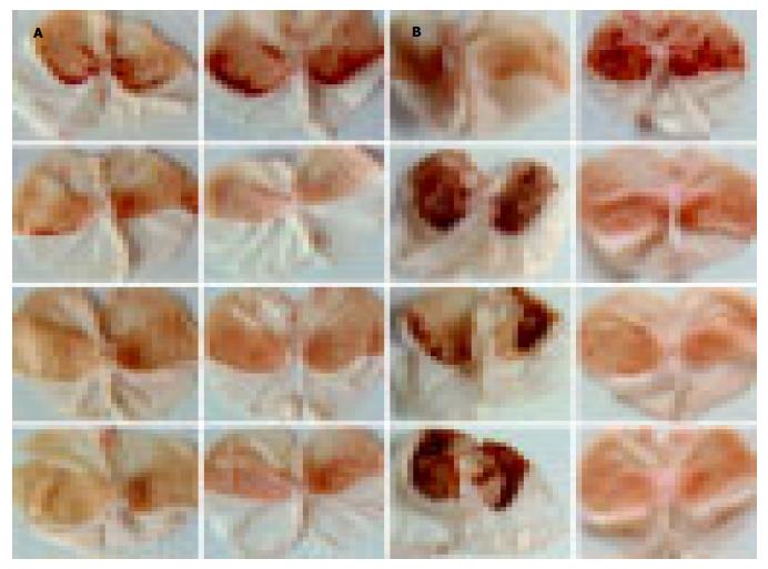
酒精性胃溃疡模型, 胃黏膜出血较严重, 有10倍镜下才能观察到的呈针尖状的出血点, 有肉眼可见的点状或线状出血灶, 更多是连成片的出血面(图1). 点状、线状出血灶及连成片的出血面, 其组织损伤达黏膜下层.
图像积分法、称质量法与记分法计量溃疡结果在不同的分级组之间均有差异, 而10倍镜下针尖状的出血点数则没有差异, 如果溃疡出血是肉眼可见的, 最好采用图像积分法、称质量法、记分法或分级法(表1).
| 分级 | n | 10倍镜下出血点数 | 记分指数 | 图像积分法溃疡面积比(%) | 称质量法溃疡面积比(%) |
| 4 | 6 | 146.2±64.9 | 84.0±27.8 | 31.57±0.16 | 40.74±0.26 |
| 2 | 5 | 197.8±63.7 | 19.6± 8.1 | 4.360±0.02 | 4.22±0.01 |
| 1 | 5 | 85.2±50.4 | 4.0± 1.0 | 2.430±0.02 | 1.03±0.01 |
图像积分法, 记分指数为111的两只小鼠, 溃疡面积比相差20.77, 记分指数为90的两只小鼠, 溃疡面积比相差33.78. 称质量法则记分指数为111的两只小鼠, 溃疡面积比相差51.38, 记分指数为90的两只小鼠, 溃疡面积比相差35.91. 这是由于4级溃疡出血较严重, 出血灶多连成片状, 用记分法主要是对点状或线状出血灶记分, 当出血灶连成片状时, 则不好掌握, 因而出现统计结果不准确. 所以, 溃疡出血较严重时, 用图像积分法及称质量法较好. 另外图像积分法, 图片大小设置为20 cm×15 cm, 比实物等比图片的放大了25倍, RGB格式, 分辨率为72像素/英寸, 加均匀的网格线, 在此放大倍数下非常清晰的记数溃疡所占的格数, 比肉眼下直接记分精确的多, 更能准确的表示溃疡出血情况, 因而图像积分法对各个分级的溃疡面积计算都适宜, 并且准确、达到测算结果的量化. 每只小鼠用称质量法与图像积分法算出的溃疡面积比之差分别是(1)38.02, (2)0.69, (3)4.89, (4)7.41, (5)1.26, (6)2.76, 由此可见, 除一只小鼠之外, 其他的差值均较小, 说明二者测量、计算结果基本相同, 并且, 称质量法测算的溃疡面积比略大于图像积分法, 这是由于溃疡部位水肿, 高出周边部, 在照片上溃疡部位周围有阴影, 在剪溃疡部位时, 可能将阴影部一同剪下称质量, 而造成误差. 同是按分级法测算所得的溃疡4级的6只小鼠, 用称质量法或图像积分法算出的溃疡面积比各只之间相差较大, 称质量法最大的相差69.91, 图像积分法最大的相差40.85, 由此可见, 分级法对溃疡病理变化程度的评定, 只能做一大体的评定标准; 而称质量法与图像积分法则能对其精确的测算与评定, 用此两种方法为评定标准, 可使实验结果更精确, 可靠.
造模Ⅰ组与造模Ⅱ组比较, 记分指数、分级数、10倍镜下出血点记数, 均无显著差异. 图像积分法、称质量法的溃疡面积比, 有显著差异. 由此可见, (1)图像积分法与称质量法是溃疡病理变化程度评定指标中较为灵敏的一个指标, 精确度高, (2)造模Ⅱ组比造模Ⅰ组造模效果好(表2).
| 组别 | 记分指数 | 分级数 | 10倍镜下出血点记数 | 图像积分法溃疡面积比(%) | 称质量法溃疡面积比(%) |
| 造模Ⅰ组 | 25.4±30.0 | 2.12±1.36 | 121.4±62.2 | 6.56±0.07 | 6.14±0.08 |
| 造模Ⅱ组 | 52.4±46.4 | 2.88±1.25 | 165.1±73.6 | 21.22±0.21 | 27.64±0.31 |
| 空白对照组 | 0 | 0 | 6.4± 5.4 | 0 | 0 |
本实验结果: 造模Ⅱ组造模效果明显比造模Ⅰ组好. 造模Ⅰ组灌服的自来水30 min后, 还没有排空, 当灌服500 mL/L乙醇造模时, 稀释了造摸用酒精浓度, 造成造模效果差, 因而, 具体应用时选造模Ⅱ组的方法较好. 通过两种轻重不同的造模方式, 造成不同分级的溃疡模型, 利于本实验不同级别溃疡结果的计量比较. 从实验结果看, 图像积分法、称质量法与记分法计量溃疡结果在不同的分级级别之间均有差异, 10倍镜下针尖状的出血点数则没有差异, 因而, 笔者认为图像积分法、称质量法、记分法及分级法均可作为溃疡结果评定的指标, 而10倍镜下针尖状的出血点数不是一个很好的指标. 4级溃疡结果统计以及造模Ⅰ组与造模Ⅱ组造模结果比较显示, 图像积分法、称质量法灵敏度和精确度优于分级法、记分法. 图像积分法与称质量法是统计溃疡面积/胃总面积的比值, 可以消除胃面积大小不均造成的误差. 对溃疡出血较轻、分级在2级以下用记分法较好, 因2级以下溃疡出血主要为点状或线状出血灶. 4级以上更多是连成片的出血面, 则以称质量法较为准确、方便, 图像积分法则对任何分级组都能准确的计量.
图像积分法与称质量法计量溃疡面积, 使溃疡面积的测量达到量化, 利用溃疡面积比对溃疡的程度进行统计分析, 使统计结果更精确, 量化. 并且, 这两种方法简单方便, 图像积分法是利用大众性的photoshop软件处理图像, 一般实验室能达到此方法所需的条件及技术要求; 称质量法则是将每张图片设置为9 cm×7 cm, A4纸打印出来(或5寸彩照), 将溃疡部剪下称质量即可, 方法简单易行, 基层实验室均能达到此要求. 因此两种方法实用性强、实用面广. 另外, 此方法还可用于肠溃疡等面积计量, 也可以对临床内窥镜检测溃疡结果进行统计学分析.
编辑: 潘伯荣 审读:张海宁
| 2. | Liu XM, Zakaria MN, Islam MW, Radhakrishnan R, Ismail A, Chen HB, Chan K, Al-Attas A. Anti-inflammatory and anti-ulcer activity of Calligonum comosum in rats. Fitoterapia. 2001;72:487-491. [PubMed] [DOI] |
| 3. | Guaraldo L, Sertie JA, Bacchi EM. Antiulcer action of the hydroalcoholic extract and fractions of Dailla rugosa Poiret in the rat. J Ethnopharmacol. 2001;76:191195. [PubMed] [DOI] |
| 4. | Yesilada E, Takaishi Y, Fujita T, Sezik E. Anti-ulcerogenic effects of Spartium junceum flowers on in vivo test models in rats. J Ethnopharmacol. 2000;70:219226. [PubMed] [DOI] |
| 5. | Tan PV, Dimo T, Dongo E. Effects of methanol, cyclohexane and methylene chlorideextracts of Bidens pilosa on various gastric ulcer models in rats. J Ethnopharmacol. 2000;73:415421. [PubMed] [DOI] |
| 6. | Xing J, Yang B, Dong Y, Wang B, Wang J, Kallio HP. Effects of sea buckthorn (Hippophae rhamnoides L.) seed and pulp oils on experimental models of gastric ulcer in rats. Fitoterapia. 2002;73:644-650. [PubMed] [DOI] |
| 7. | Abdel-Salam OM, Baiuomy AR, El-batran S, Arbid MS. Evaluation of the anti-inflammatory, anti-nociceptive and gastric effects of Ginkgo biloba in the rat. Pharmacol Res. 2004;49:133142. [PubMed] [DOI] |
| 8. | Grover JK, Adiga G, Vats V, Rathi SS. Extracts of Benincasa hispida prevent development of experimental ulcers. J Ethnopharmacol. 2001;78:159-164. [PubMed] [DOI] |
| 9. | Rodriguez JA, Astudillo L, Schmeda-Hirschmann G. Oleanolic acid promotes healing of acetic acid-induced chronic gastric lesions in rats. Pharmacol Res. 2003;48:291-294. [PubMed] [DOI] |
| 10. | Jiang P, Chang L, Pan CS, Qi YF, Tang CS. Protective role of metallothionein in stress-induced gastric ulcer in rats. World J Gastroenterol. 2005;11:2739-2743. [PubMed] [DOI] |
| 11. | Wang L, Hu CP, Deng PY, Shen SS, Zhu HQ, Ding JS, Tan GS, Li YJ. The protective effects of rutaecarpine on gastric mucosa injury in rats. Planta Med. 2005;71:416-419. [PubMed] [DOI] |
| 12. | Zhang JF, Zhang YM, Yan CD, Zhou XP. Neuroregulative mechanism of hypothalamic paraventricular nucleus on gastric ischemia-reperfusion injury in rats. Life Sci. 2002;71:1501-1510. [PubMed] [DOI] |
| 13. | Zhou XP, Zhang JF, Yan CD, Zhang YM. Effects of electrical stimulation of lateral hypothalamic area on gastric ischemia-reperfusion injury in rats. Shengli Xuebao. 2002;54:435-440. [PubMed] |
| 14. | Zhang JF, Zhang YM, Yan CD, Zhou XP, Qi YJ. Protective effects of paraventricular nucleus stimulation and vasopressin on gastric ischemia-reperfusion injury in rats. Shengli Xuebao. 2002;54:133-138. [PubMed] |
| 15. | Villegas I, Alarcon de la Lastra C, La Casa C, Motilva V, Martin MJ. Effects of food intake and oxidative stress on intestinal lesions caused by meloxicam and piroxicam in rats. Eur J Pharmacol. 2001;414:79-86. [PubMed] [DOI] |
| 16. | Sartori NT, Canepelle D, de Sousa PT Jr, Martins DT. Gastroprotective effect from Calophyllum brasiliense Camb. bark on experimental gastric lesions in rats and mice. J Ethnopharmacol. 1999;67:149-156. [PubMed] [DOI] |
| 17. | Chiu PJ, Gerhart C, Brown AD, Barnett A. Effects of a gastric antisecretory-cytoprotectant 2-methyl-8- (phenylmethoxy) imidazo[1, 2-a]pyridine-3-acetonitrile (Sch 28 080) on cysteamine, reserpine and stress ulcers in rats. Arzneimittelforschung. 1984;34:783-786. [PubMed] |
| 18. | Robert A, Nezamis JE, Lancaster C, Davis JP, Field SO, Hanchar AJ. Mild irritants prevent gastric necrosis through "adaptive cytoprotection" mediated by prostaglandins. Am J Physiol. 1983;245:113-121. [PubMed] |
| 19. | Paiva LA, Rao VS, Gramosa NV, Silveira ER. Gastroprotective effect of Copaifera langsdorffii oleo-resin on experimental gastric ulcer models in rats. J Ethnopharmacol. 1998;62:73-78. [PubMed] [DOI] |
| 20. | Uchida M, Takayama M, Kato Y, Tsuchiya S, Horie S, Watanabe K. A novel method to produce extensive gastric antral ulcer in rats: pharmacological factors involved in the etiology of antral ulceration. J Physiol Paris. 1999;93:437-442. [PubMed] [DOI] |


