修回日期: 2004-11-15
接受日期: 2004-11-22
在线出版日期: 2005-01-01
目的: 应用蛋白质组学技术分析高低转移表型大肠癌SW480细胞株和LoVo细胞株间的蛋白质表达谱差异.
方法: 以双向电泳技术分离两种细胞株总蛋白质, 银染显色, 进行差异蛋白质分析.
结果: SW480和LoVo细胞株双向电泳图谱蛋白质点数分别为1184±47和1124±54, 共获得88±5的蛋白质差异点, 其中48±3个点仅在SW480细胞株中表达或表达明显增强, 41±3个点仅在LoVo细胞中表达或表达明显增强.
结论: 高低转移表型大肠癌SW480细胞株和LoVo细胞株间的蛋白质表达谱存在一定的差异, 对这些差异点进行鉴定将为研究大肠癌转移机制提供一定的线索.
引文著录: 童华生, 张亚历, 姜泊, 赖卓胜, 马文敏, 耿焱. 高低转移表型大肠癌细胞株蛋白质表达谱差异初步分析. 世界华人消化杂志 2005; 13(1): 109-112
Revised: November 15, 2004
Accepted: November 22, 2004
Published online: January 1, 2005
N/A
- Citation: N/A. N/A. Shijie Huaren Xiaohua Zazhi 2005; 13(1): 109-112
- URL: https://www.wjgnet.com/1009-3079/full/v13/i1/109.htm
- DOI: https://dx.doi.org/10.11569/wcjd.v13.i1.109
大肠癌是消化系统的一种常见肿瘤, 死亡率高, 主要与其广泛转移有关, 故此大肠癌的转移机制成为研究的热点, 而研究不同转移潜能的大肠癌细胞株间的分子表达谱差异可为其提供线索. 大肠癌SW480细胞株为一种原发的大肠腺癌, 具有低转移潜能[1-5], 而LoVo细胞株来源于结肠腺癌的左锁骨上转移结节[6], 是一种具有高转移潜能的细胞株[7-12], 由于二者转移表型上存在着差异, 可作为研究肿瘤转移的细胞模型.
目前关于大肠癌转移分子机制有基因表达谱差异的报道, 而蛋白质研究方面多为强调单个或少数几个分子的作用[13-15], 缺乏蛋白质水平的系统性、整体性研究. 近年蛋白质组学(proteomics)在探寻肿瘤早期发现和诊断肿瘤生物标记和新的抗肿瘤药靶, 评价药物疗效和毒性方面取得了进展, 为我们研究肿瘤转移提供了思路[16]. 鉴于此, 我们采用蛋白质组学技术分析SW480和LoVo细胞株蛋白质表达谱, 对其差异作初步分析.
人大肠癌SW480(ATCC编号: CCL-228)细胞株和LoVo(ATCC编号: CCL-229)细胞株均来自ATCC(美国组织培养保藏中心), 由上海生物细胞所提供. 两细胞株均用100 mL/L小牛血清(fetal calf serum, FCS)+RPMI1640培养液进行传代培养. IPG干胶条pH4-7与相应的IEF buffer及三丁基磷(TBP)购自Bio-Rad公司; 超纯尿素、硫脲、碘乙酰胺、二硫苏糖醇(DTT)、甘油、亚甲叉双丙烯酰胺、四甲基乙二胺(TEMED)、矿物油、过硫酸胺和溴酚蓝均购自Sigma公司; 丙烯酰胺(和苯甲基磺酰氟(PMSF)购自BEBCO公司; 三羟甲基胺基甲烷(Tris)购自Genview公司; CHAPS购自Amresco公司; 其余试剂均为分析纯以上. 主要的仪器有Bio-Rad公司的PROTEAN IEF cell系统和Bio-Rad PROTEANⅡ xi系统.
1.2.1 样品的制备: 细胞培养于100 mL/L FCS+RPMI1640培养基中, 待生长至对数生长期密度约为80%时用细胞括括下细胞离心收集. 按细胞/裂解液体积比1:10加入细胞裂解液(7 mol/L尿素, 2 mol/L硫脲, 40 g/LCHAPS, 65 mmol/L DTT), 同时加入1 mmol/L苯甲基磺酰氟(Phenylmethylsufosyl, PMSF), 旋涡振荡, 反复液氮快速冻融4次, 加DnaseⅠ50 mg/L和Rnase A 25 mg/L室温作用15 min, 4 ℃ 15000 g离心1 h, 收集上清液-80 ℃冻存备用, Bradford法测定蛋白浓度[17].
1.2.2 第一向固相pH梯度等电聚焦: 略加改动Gorg et al[18]方法, 蛋白质样品250 μg加入重水化液(7 mol/L尿素, 2 mol/L硫脲, 20 g/L CHAPS, 50 mmol/L DTT)至终体积300 μL, 临时加入2 mmol/L TBP和2 mL/L IEF buffer, 室温充分溶解. 然后移入胶条槽中, 胶面朝下放入17 cm固相pH梯度(immobilized pH gradient, IPG)干胶条, 避免产生气泡, 1 h后覆盖3 mL矿物油, 20 ℃重水化12 h. 取出重水化胶条转移至等电聚焦(isoelectric focusing, IEF)聚焦盘槽中, 加适量的矿物油后进行IEF. IEF条件: 250V 1/2 h→500V 1 h→1000V 1 h→5000V 3 h→10000V 70000Vh→500V hold, 温度为20 ℃, 最高电流为50 μA.
1.2.3 胶条平衡: 胶条依次在平衡液Ⅰ(5 mol/L尿素, 20 g/L SDS, 50 mmol/Ltris-HCl, 20%甘油, 2%DTT)和平衡液Ⅱ(5 mol/L尿素, 20 g/L SDS, 50 mmol/L Tris-HCl, 200 mL/L甘油, 25 g/L碘乙酰胺)各5 mL中平衡10 min.
1.2.4 二向垂直SDS-PAGE: 将平衡后的IPG胶条转移至13%凝胶的上方, 胶条一端置低分子量蛋白质标准, 5g/L的琼脂糖封闭[19]. 电泳参数如下: 12mA/胶1/2 h, 24mA/胶直至溴酚蓝前沿抵达凝胶底部为止.
1.2.5 银染及图像可视化: 银染步骤按以下程序进行: 固定→浸泡→漂洗→银染→显色→终止. 通过PowerLook 1100扫描仪获取图像, 用Melanine3分析软件对图像进行分析.
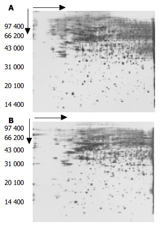
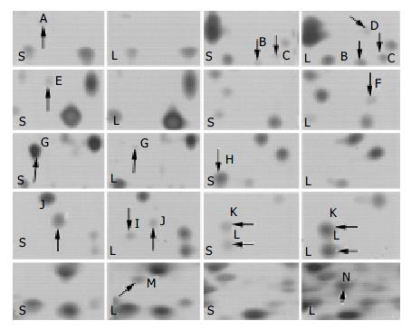
相同的实验条件和参数设置的情况下, 实验重复三次, 蛋白质上样量均为250 μg, 测得SW480细胞株和LoVo细胞株双向电泳(two-dimensional electrophoresis, 2-DE)图谱蛋白质斑点数分别为1184±47和1124±54. 图1为两细胞株的2-DE图, 从中可看出两细胞株的蛋白质表达谱分布相似. 经对两细胞株2-DE图谱进行匹配分析, 有88±5的蛋白质差异点, 其中48±3个点仅在SW480细胞株中表达或表达明显增强, 41±3个点仅在LoVo细胞中表达或表达明显增强. 图2为部分差异表达点局部放大示为意图.
大肠癌是全球的第四大恶性肿瘤, 其死亡率与肿瘤分级关系密切, 大约有95%以上大肠癌转移患者是不可治愈的[20], 因此研究大肠癌转移成为该领域中重要的环节之一. 目前基因组学技术已运用于大肠癌转移分子机制研究, 并且发现了一系列的基因表达谱的差异[21-23], 但基因与蛋白质的线性关系并非完全一致[24]. 因此, 无论是从基因组的局限性, 还是从蛋白质自身发展的需要, 大规模、全方位的蛋白质研究势在必行[25-26]. 1994年, Williams和澳大利亚悉尼Maquarie大学的Wilkins et al首先提出了"蛋白质组(proteome)"名词[27-28], 其含义是指一个基因组、一个细胞或组织内表达的全部蛋白质. 蛋白质组学(proteomics)则是一衍生概念, 其宗旨为通过对蛋白质的丰度和性质变化的考察, 揭示蛋白质的活动规律, 解释基因调控机制, 从而透视到生命的本质[29]. 基于此, 我们采用蛋白质组学技术对不同转移潜能大肠癌细胞株SW480和LoVo进行蛋白质表达谱差异分析.
从我们的实验结果来看, 两种细胞株双向电泳图谱中多数蛋白质的分布和表达量是一致的, 这表明SW480和LoVo细胞株具有大肠癌的共同蛋白质表达谱, 提示二者有大肠癌共有特性. 但是经过分析我们发现有88±5的蛋白质差异点, 其中48±4个点仅在SW480细胞株中表达或表达明显增强, 41±3个点仅在LoVo细胞中表达或表达明显增强, 这些差异蛋白质可能在大肠癌的转移中发挥一定的作用. 我们推测一些涉及肿瘤转移过程各个阶段的蛋白质可能存在表达上的差异, 实验尚待下一步证实.
目前作为蛋白质组学的支撑技术-双向电泳的分辩率尚不能满足人类基因组编码的全部蛋白质(约5万个)[30], 但应用该技术初步表明不同转移潜能的大肠癌细胞株蛋白质表达谱存在着一定的差异. 欲鉴定出这些差异蛋白质, 尚须进行N端测序、氨基酸组成分析、质谱分析、肽质谱及数据库搜寻等工作[31]. 我们应注意到, 用银染的蛋白质进行鉴定有一定的困难存在, 而且对于一些低丰度差异蛋白质的鉴定仍是蛋白质组学的瓶颈. 由于这些低丰度蛋白质往往具有重要的生物学功能, 其鉴定失败将有可能意味着大肠癌转移重要相关分子的丢失. 尽管如些, 蛋白质组学技术毕竟在蛋白质整体水平研究方向迈出了重要的一步, 相信随着该技术的逐步完善, 我们将进一步认识大肠癌转移这一生物现象.
蛋白质双向电泳实验内容在南方医科大学肿瘤研究所完成, 在此感谢给予的帮助和指导.
编辑: 张海宁
| 1. | Leibovitz A, Stinson JC, McCombs WB, McCoy CE, Mazur KC, Mabry ND. Classification of human colorectal adenocarcinoma cell lines. Cancer Res. 1976;36:4562-4569. [PubMed] |
| 2. | Liang L, Ding YQ, Li X, Yang GZ, Xiao J, Lu LC, Zhang JH. Construction of a metastasis-associated gene subtracted cDNA library of human colorectal carcinoma by suppression subtraction hybridization. World J Gastroenterol. 2004;10:1301-1305. [PubMed] |
| 3. | Hewitt RE, McMarlin A, Kleiner D, Wersto R, Martin P, Tsokos M, Stamp GW, Stetler-Stevenson WG. Validation of a model of colon cancer progression. J Pathol. 2000;192:446-454. [PubMed] |
| 4. | Mc Donnell S, Chaudhry V, Mansilla-Soto J, Zeng ZS, Shu WP, Guillem JG. Metastatic and non-metastatic colorectal cancer (CRC) cells induce host metalloproteinase production in vivo. Clin Exp Metastasis. 1999;17:341-349. [PubMed] |
| 5. | Lin JC, Cheng JY, Tzeng CC, Yeh MY, Meng CL. An animal model for colon cancer metastatic cell line with enhanced metastasizing ability. Establishment and characterization. Dis Colon Rectum. 1991;34:458-463. [PubMed] |
| 6. | Drewinko B, Romsdahl MM, Yang LY, Ahearn MJ, Trujillo JM. Establishment of a human carcinoembryonic antigen-producing colon adenocarcinoma cell line. Cancer Res. 1976;36:467-475. [PubMed] |
| 8. | 李 小卫, 丁 彦青, 蔡 俊杰, 张 素娟. 唾液酸化的路易斯寡糖-X抗原在结直肠癌细胞系LoVo、HT29中的表达及其与转移潜能的相关性. 中华病理学杂志. 2000;29:119-122. |
| 10. | Kondo Y, Arii S, Mori A, Furutani M, Chiba T, Imamura M. Enhancement of angiogenesis, tumor growth, and metastasis by transfection of vascular endothelial growth factor into LoVo human colon cancer cell line. Clin Cancer Res. 2000;6:622-630. [PubMed] |
| 11. | Daémi N, Vallet T, Thomasset N, Jacquier MF, Zebda N, Doré JF, Sordat B, Rémy L. Expression of the alpha 6, beta 1 and beta 4 integrin subunits, basement membrane organization and proteolytic capacities in low and high metastatic human colon carcinoma xenografts. Invasion Metastasis. 1995;15:103-115. [PubMed] |
| 12. | Liu L, Wu DH, Li ZG, Yang GZ, Ding YQ. Effects of KAI1/CD82 on biological behavior of human colorectal carcinoma cell line. World J Gastroenterol. 2003;9:1231-1236. [PubMed] |
| 13. | Wang WS, Chen PM, Hsiao HL, Wang HS, Liang WY, Su Y. Overexpression of the thymosin beta-4 gene is associated with increased invasion of SW480 colon carcinoma cells and the distant metastasis of human colorectal carcinoma. Oncogene. 2004;23:6666-6671. [PubMed] |
| 14. | Baldus SE, Mönig SP, Huxel S, Landsberg S, Hanisch FG, Engelmann K, Schneider PM, Thiele J, Hölscher AH, Dienes HP. MUC1 and nuclear beta-catenin are coexpressed at the invasion front of colorectal carcinomas and are both correlated with tumor prognosis. Clin Cancer Res. 2004;10:2790-2796. [PubMed] |
| 15. | de Albuquerque Garcia Redondo P, Nakamura CV, de Souza W, Morgado-Díaz JA. Differential expression of sialic acid and N-acetylgalactosamine residues on the cell surface of intestinal epithelial cells according to normal or metastatic potential. J Histochem Cytochem. 2004;52:629-640. [PubMed] |
| 16. | Wu W, Hu W, Kavanagh JJ. Proteomics in cancer research. Int J Gynecol Cancer. 2002;12:409-423. [PubMed] |
| 17. | Blomberg A, Blomberg L, Norbeck J, Fey SJ, Larsen PM, Larsen M, Roepstorff P, Degand H, Boutry M, Posch A. Interlaboratory reproducibility of yeast protein patterns analyzed by immobilized pH gradient two-dimensional gel electrophoresis. Electrophoresis. 1995;16:1935-1945. [PubMed] |
| 18. | Görg A, Obermaier C, Boguth G, Harder A, Scheibe B, Wildgruber R, Weiss W. The current state of two-dimensional electrophoresis with immobilized pH gradients. Electrophoresis. 2000;21:1037-1053. [PubMed] |
| 19. | Görg A, Obermaier C, Boguth G, Csordas A, Diaz JJ, Madjar JJ. Very alkaline immobilized pH gradients for two-dimensional electrophoresis of ribosomal and nuclear proteins. Electrophoresis. 1997;18:328-337. [PubMed] |
| 20. | Lars P. Colorectal cancer. Cabriele BP主编. Gastroenterology and Hepatology. 第1版. 西安: 世界图书出版社 2000; 351-364. |
| 21. | Weng DS, Ding YQ, Sun Q. Differences in gene expression profile between high and low metastatic human colorectal cancer cell lines. Diyi Junyi Daxue Xuebao. 2004;24:256-259. [PubMed] |
| 22. | Li M, Lin YM, Hasegawa S, Shimokawa T, Murata K, Kameyama M, Ishikawa O, Katagiri T, Tsunoda T, Nakamura Y. Genes associated with liver metastasis of colon cancer, identified by genome-wide cDNA microarray. Int J Oncol. 2004;24:305-312. [PubMed] |
| 23. | Yeatman TJ, Cher ML, Mao W, Wloch M, Tedesco T. Identification of genetic alterations associated with the process of human experimental colon cancer liver metastasis in the nude mouse. Clin Exp Metastasis. 1996;14:246-252. [PubMed] |
| 24. | Anderson L, Seilhamer J. A comparison of selected mRNA and protein abundances in humor liver. Electrophoresis. 1997;18:533-537. |
| 27. | Blackstock WP, Weir MP. Proteomics: quantitative and physical mapping of cellular proteins. Trends Biotechnol. 1999;17:121-127. [PubMed] |
| 28. | Wasinger VC, Cordwell SJ, Cerpa-Poljak A, Yan JX, Gooley AA, Wilkins MR, Duncan MW, Harris R, Williams KL, Humphery-Smith I. Progress with gene-product mapping of the Mollicutes: Mycoplasma genitalium. Electrophoresis. 1995;16:1090-1094. [PubMed] |
| 29. | Anderson NL, Anderson NG. Proteome and proteomics: new technologies, new concepts, and new words. Electrophoresis. 1998;19:1853-1861. [PubMed] |


