修回日期: 2003-09-05
接受日期: 2003-09-24
在线出版日期: 2004-03-15
目的: 探讨胃癌中VEGF, Flt1, bFGF, P53的表达及意义.
方法: 应用SABC免疫组化法, 研究VEGF, Flt1, bFGF, P53在胃癌中表达及与胃癌生长转移、临床病理特征、预后的关系.
结果: 胃癌VEGF表达与肿瘤浸润深度有关(浆膜、浆膜外 vs 肌层和黏膜层, P<0.01). P53表达与淋巴结转移相关(P53阳性淋巴结转移组 vs P53阴性淋巴结无转移组, P<0.05).VEGF与Flt1表达呈正相关(VEGF表达在Flt1阳性组 vs 在Flt1阴性组, P<0.01); 影响胃癌预后的因素有临床病理分期、VEGF表达、肿瘤浸润深度、手术方式. Kaplan-Meier研究显示VEGF表达与胃癌生存预后相关(P<0.05).Flt1, bFGF, P53表达与生存预后无关(P>0.05).
结论: P53表达与淋巴结转移相关. VEGF表达与肿瘤浸润深度和胃癌生存预后相关, VEGF表达可作为预测胃癌预后一项很好的指标.
引文著录: 段伦喜, 钟德午, 胡辅珍, 赵华, 杨竹林, 易文君, 舒国顺, 华颂文. 胃癌组织VEGF, Flt1, bFGF, P53表达与胃癌预后的关系. 世界华人消化杂志 2004; 12(3): 546-549
Revised: September 5, 2003
Accepted: September 24, 2003
Published online: March 15, 2004
AIM: To investigate the relationship between the expression of VEGF, Flt1, bFGF and P53, the clinicopathological characteristics and outcome in patients with gastric carcinoma.
METHODS: The relationship between VEGF, Flt1, bFGF, P53 expression and clinicopathological characteristics and outcome in the patient was assessed by streptoavidin-biotin method of immunohistochemistry with polyclonal antibodies against VEGF, Flt1, bFGF, and P53 protein. The survival curves were formulated using Kaplan-Meier method and analyzed by the log-rank test, and the influence of each variable on suvival was assessed by the Cox' s proportional hazard model.
RESULTS: VEGF expression was closely correlated with serosal invasion (Se, Sei invasion vs Pm, SS and M, SM invasion, P < 0.01). Expression of P53 was obviously higher in the patients with lymph node metastasis than those without (lymph node metastasis vs non-lymph node metastasis, P < 0.05). There was a positive correlation between VEGF and Flt1 expression (VEGF expression in Flt1 positive group vs Flt1 negative group, P < 0.01). The factors that affected the prognosis in patients with gastric carcinoma were PTNM stage, VEGF expression, serosal invasion, and surgical curability. Flt1, bFGF, and P53 expression had no influence on the prognosis of patients with gastric carcinoma (P > 0.05).
CONCLUSION: P53 expression has significant relationship with lymph node metastasis in gastric carcinoma. VEGF expression is correlated with serosal invasion and the prognosis and may be a good prognostic indicator in gastric carcinoma.
- Citation: Duan LX, Zhong DW, Hu FZ, Zhao H, Yang ZL, Yi WJ, Shu GS, Hua SW. Relationship between expression of VEGF, Flt1, bFGF and P53and outcome in patients with gastric carcinoma. Shijie Huaren Xiaohua Zazhi 2004; 12(3): 546-549
- URL: https://www.wjgnet.com/1009-3079/full/v12/i3/546.htm
- DOI: https://dx.doi.org/10.11569/wcjd.v12.i3.546
胃癌是最常见癌肿之一[1-6], 探讨胃癌生物学行为及预后相关因素有重要意义[7-10] . 实体肿瘤的生长与转移依赖于血管生成[11]. 肿瘤的血管生成赖于多种相关因子的诱导和调节[12], 在众多与其有关的因子中, VEGF (vascular endothelial growth factor, VEGF)是迄今鉴定出来的最重要的血管生成因子.我们探讨胃癌中VEGF及其受体Flt1和P53, bFGF的表达与预后的关系.
1994-01/1996-10本院普外科收治胃癌52例.年龄28-73, (平均51); 男34例, 女18例; 临床病理分期(按IUCC1988年标准)Ⅰ级14例, Ⅱ级9例, Ⅲ级15例, Ⅳ级14例; 根治性手术39例, 姑息性手术13例. 所有病例术前未作放疗和化疗, 术后均接受随访, 大于5年者40例. 5年内死亡25例, 其中根治手术13例, 姑息手术12例; 生存15例, 均为根治手术.羊抗人FGF-21(147)-G(IgG)(1: 200), P53(FL-393)-G(IgG)(1: 200), 兔抗人VEGF(147)(IgG)(1: 200), Flt1(C-17)(IgG)(1: 200)多克隆抗体, SABC试剂盒均购于武汉博士德公司.
采用SABC法, 以已知阳性大肠癌切片作为阳性对照, 以PBS替代一抗作为阴性对照. 染色步骤: 切片脱蜡, 30 ml/L H2O2甲醇液封闭内源性过氧化酶, 消化液消化, 滴加一抗、二抗和SABC试剂, AEC显色, 苏木精复染, 梯度酒精脱水, 二甲苯透明、中性树胶封固. 显微镜下双盲法阅片, 在细胞核或细胞质内、细胞膜被染成红色者为阳性细胞. (1) VEGF, P53蛋白: 阳性癌细胞 大于或等于10%为阳性, 无阳性细胞或阳性癌细胞小于10%为阴性[13-14]; (2)Flt1蛋白: 肿瘤血管内皮细胞阳性染色判为阳性, 肿瘤血管内皮细胞阴性染色判为阴性; (3)bFGF: 以胞质胞膜被染成红色者为阳性细胞, 采用图像分析系统(HPIAS-1000P)对染色强度进行定量, 按强度不同分为0到+++, 以0代表未见染色, +++代表最强染色.
统计学处理 由Spss10.0统计软件完成, 率的比较采用采x2检验, Kaplan-Meier法作生存率曲线, Logrank法作时序检验, 各指标对生存率的影响采用Cox模型多因素分析, 检验水准P = 0.05.
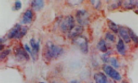
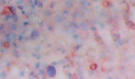
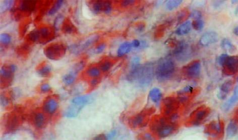
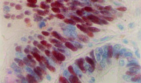
VEGF表现为细胞膜或胞质阳性(图1), 阳性表达率为53.8% (28/52). Flt1表现为胞质阳性, 以肿瘤血管内皮细胞阳性表达为主(图2), 阳性表达率为51.9% (27/52). bFGF 表现为胞质胞膜阳性, 少数肿瘤细胞核阳性(图3), 阳性表达率71.2%(37/52). P53主要为胞核阳性(图4), 阳性表达率34.6%(18/52).
VEGF表达与肿瘤浸润深度有关(P<0.01). P53表达与淋巴结转移有关(P<0.05). F1t1, bFGF表达与临床病理分期、组织学类型、浸润深度、淋巴转移、远处转移之间均无关(P>0.05), 见表1.
Flt1阳性组VEGF阳性率为74.1%, Flt1阴性组VEGF阳性率为32.0%, 二者有显著性差异(P<0.05), 表明VEGF与Flt1呈正相关. 其他各因子之间无相关性. 将52例生存时间和各因素定量化数据输入计算机得到COX回归方程为: hj(t)=ho(t)exp (0.7221临床病理分期+1.471浸润深度+1.2605VEGF表达-1.2770手术方式). 说明对胃癌预后有影响的因素只有临床病理分期、肿瘤浸润深度、VEGF表达、手术方式四因素, 其余各因素对生存预后无影响.而从优势比看, 影响程度的大小依次为: 浸润深度、手术方式、VEGF表达、临床病理分期.
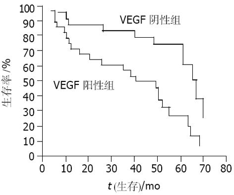
将VEGF表达分为阳性组和阴性组, 制出Kaplan-Meier生命曲线(图5), 以logrank检验法检验, 其P = 0.0030表明两组生存率有显著性差异.同样对Flt1, bFGF, P53制出生命曲线图并作logrank检验, P>0.05, 表明以Flt1, bFGF, P53蛋白表达分组, 两组生存率无显著性差异.
肿瘤生长赖于血管生成, 作为重要的血管生成因子, VEGF与胃癌的发生发展转移复发有关[14-17]. VEGF和胃癌血管生成淋巴结转移以及胃癌发展密切相关, 结合微血管计数可以预测胃癌的分期和转移[15]; VEGF血清含量增高, 预示胃癌的转移和预后不良[16]. 本研究显示VEGF表达与胃癌浸润深度有关, 与其他临床病理因素无关. 同时我们采用Kaplan-Meier法分别对VEGF, Flt1, P53阳性表达组和阴性表达组病例生存率作了生存曲线图对比, 发现VEGF与胃癌生存率和预后相关, 而Flt1, P53与胃癌生存率和预后无关. K-M法生存率分析只是一种单因素分析方法, 不能去除各因子间的混杂影响. 我们又应用Cox多因素模型对临床病理分期(PTNM分期)、细胞学分级、浸润深度、淋巴转移、远处转移、手术方式、VEGF、Flt1、P53 表达进行了多因素回归分析, 最后被引入Cox回归方程的只有: 手术方式、临床病理分期、VEGF表达和浸润深度四个因素, 表明 VEGF可判断胃癌预后, 而Flt1、P53与胃癌预后无明显关系.
VEGF通过与受体Flt1和KDR的接合而刺激肿瘤血管内皮细胞的生长, 在肿瘤的血管生成中起到非常重要的作用. 李江et al[18]研究指出肝细胞肝癌中血管形成主要是由VEGF/ Flt1系统介导的, 血管形成丰富的区域, 细胞凋亡的敏感性和发生率降低; 胃癌细胞增值和血管内皮细胞增值相互影响且机制复杂, VEGF受体在其中起到重要的作用[19-20]. 显性阴性的KDR突变体引起的肿瘤血管生成的抑制不可被Flt1恢复, 说明Flt1不介导有丝分裂作用, 可能介导VEGF引起的非有丝分裂反应.本研究 Flt1 阳性组与Flt1阴性组VEGF阳性率有显著性差异, Flt1表达随VEGF 表达上调而上调, 表明VEGF与Flt1表达有相关性, 同时Flt1不但在血管内皮细胞表达, 同时在少数肿瘤细胞上亦表达, 表明VEGF除了旁分泌机制作用血管内皮细胞外, 也存在自泌机制.
bFGF促进细胞黏膜组织修复[21], bFGF可能为血管内皮细胞有丝分裂原并参与肿瘤血管生成[22]. bFGF表达与肿瘤细胞分化、淋巴转移、临床病理分期、血管计数、pCNA指数、DNA倍体含量相关[22-23]. 本组病例bFGF表达与临床病理各因素、胃癌生存率及预后无关.表明bFGF在有些类型肿瘤中有促血管生成作用, 而在有些类型肿瘤如胃癌中无直接促血管生成作用. 陶厚权et al[24]研究指出Rg3能有效抑制人胃癌血管生成, 其机制可能与减少VEGF和bFGF表达有关.因此, bFGF在胃癌肿瘤血管生成中的作用, 有待我们进一步研究.
P53基因是一种抑癌基因, 突变型P53可导致细胞癌变, 已经证实P53蛋白表达与多种恶性肿瘤的发生发展浸润转移预后有关[25-27]. 胃幽门螺杆菌感染可诱导P53基因突变[28-29], P53过度表达又可导致肿瘤血管生成增加[17], 后者可能与通过上调VEGF蛋白表达有关[30]. 本组病例提示突变型P53表达与VEGF, Flt1, bFGF蛋白表达无关, 我们认为P53突变能否使VEGF表达上调值得进一步探讨研究. 本组病例显示突变型P53表达与淋巴结转移有关, 提示检测突变型P53表达对预测胃癌淋巴结转移可能有一定价值.
编辑: N/A
| 1. | Sun L, Wang X. Effects of allicin on both telomerase activity and apoptosis in gastric cancer SGC-7901 cells. World J Gastroenterol. 2003;9:1930-1934. [PubMed] [DOI] |
| 2. | Zhao GH, Li TC, Shi LH, Xia YB, Lu LM, Huang WB, Sun HL, Zhang YS. Relationship between inactivation of p16 gene and gastric carcinoma. World J Gastroenterol. 2003;9:905-909. [PubMed] [DOI] |
| 3. | Zhao XH, Gu SZ, Liu SX, Pan BR. Expression of estrogen receptor and estrogen receptor messenger RNA in gastric carcinoma tissues. World J Gastroenterol. 2003;9:665-669. [PubMed] [DOI] |
| 4. | Chen H, Wang LD, Guo M, Gao SG, Guo HQ, Fan ZM, Li JL. Alterations of p53 and PCNA in cancer and adjacent tissues from concurrent carcinomas of the esophagus and gastric cardia in the same patient in Linzhou, a high incidence area for esophageal cancer in northern China. World J Gastroenterol. 2003;9:16-21. [PubMed] [DOI] |
| 5. | 王 孟薇, 杨 少波, 张 子其, 祝 庆孚, 王 刚石, 李 晖, 姚 晨, 吴 本俨, 尤 纬缔. 老年人胃癌前黏膜癌变的胃镜随访. 世界华人消化杂志. 2003;11:1279-1281. [DOI] |
| 7. | Ding YB, Chen GY, Xia JG, Zang XW, Yang HY, Yang L. Association of VCAM-1 overexpression with oncogenesis, tumor angiogenesis and metastasis of gastric carcinoma. World J Gastroenterol. 2003;9:1409-1414. [PubMed] [DOI] |
| 8. | Li HX, Chang XM, Song ZJ, He SX. Correlation between expression of cyclooxygenase-2 and angiogenesis in human gastric adenocarcinoma. World J Gastroenterol. 2003;9:674-677. [PubMed] [DOI] |
| 9. | Shi H, Xu JM, Hu NZ, Xie HJ. Prognostic significance of expression of cyclooxygenase-2 and vascular endothelial growth factor in human gastric carcinoma. World J Gastroenterol. 2003;9:1421-1426. [PubMed] [DOI] |
| 10. | Yang L, Kuang LG, Zheng HC, Li JY, Wu DY, Zhang SM, Xin Y. PTEN encoding product: a marker for tumorigenesis and progression of gastric carcinoma. World J Gastroenterol. 2003;9:35-39. [PubMed] [DOI] |
| 14. | Feng CW, Wang LD, Jiao LH, Liu B, Zheng S, Xie XJ. Expression of P53, inducible nitric oxide synthase and vascular endothelial growth factor in gastric precancerous and cancerous lesions: correlation with clinical features. BMC Cancer. 2002;2:1471-2407. [PubMed] [DOI] |
| 15. | Song ZJ, Gong P, Wu YE. Relationship between the expression of iNOS, VEGF, tumor angiogenesis and gastric cancer. World J Gastroenterol. 2002;8:591-595. [PubMed] [DOI] |
| 17. | Maehara Y, Kabashima A, Koga T, Tokunaga E, Takeuchi H, Kakeji Y, Sugimachi K. Vascular invasion and potential for tumor angiogenesis and metastasis in gastric carcinoma. Surgery. 2000;128:408-416. [PubMed] [DOI] |
| 19. | Zhang H, Wu J, Meng L, Shou CC. Expression of vascular endothelial growth factor and its receptors KDR and Flt-1 in gastric cancer cells. World J Gastroenterol. 2002;8:994-998. [PubMed] [DOI] |
| 20. | Ren J, Dong L, Xu CB, Pan BR. The role of KDR in the interactions between human gastric carcinoma cell and vascular endothelial cell. World J Gastroenterol. 2002;8:596-601. [PubMed] [DOI] |
| 21. | Fu XB, Yang YH, Sun TZ, Gu XM, Jiang LX, Sun XQ, Sheng ZY. Effect of intestinal ischemia-reperfusion on expressions of endogenous basic fibroblast growth factor and transforming growth factor in lung and its relation with lung repair. World J Gastroenterol. 2000;6:353-355. [PubMed] [DOI] |
| 22. | Yamaguchi K, Ura H, Yasoshima T, Shishido T, Denno R, Hirata K. Liver metastatic model for human gastric cancer established by orthotopic tumor cell implantation. World J Surg. 2001;25:131-137. [PubMed] [DOI] |
| 25. | Xu AG, Li SG, Liu JH, Gan AH. The function of apoptosis and protein expression of bcl-2, P53 and C-myc in the development of gastric cancer. World J Gastroenterol. 2000;6:27. [PubMed] |
| 26. | Smith DR, Ji CY, Goh HS. Prognostic significance of p53 overexpression and mutation in colorectal adenocarcinomas. Br J Cancer. 1996;74:216-223. [PubMed] [DOI] |
| 27. | Niu ZS, Li BK, Wang M. Expression of p53 and c-myc genes and its clinical relevance in the hepatocellular carcinomatous and pericarcinomatous tissuses. World J Gastroenterol. 2002;8:822-826. [PubMed] [DOI] |
| 28. | Gao HJ, Yu LZ, Bai JF, Peng YS, Sun G, Zhao HL, Miu K, Lu XZ, Zhang XY, Zhao ZQ. Multiple genetic alterations and behavior of cellular biology in gastric cancer and other gastric mucosal lesions: H pylori infection, histological types and staging. World J Gastroenterol. 2000;6:848-854. [PubMed] [DOI] |


