修回日期: 2002-11-20
接受日期: 2002-12-02
在线出版日期: 2003-08-15
研究美蓝染色后用放大电子结肠镜观察结肠息肉的微细结构类型与组织病理学的关系.
使用0.5%美蓝对电子结肠镜检查到有息肉的部位进行息肉及邻近的黏膜进行染色, 然后用放大电子结肠镜观察. 按Kudo's标准记录正常结肠黏膜和息肉, 然后行活检或息肉电切术, 以便组织病理学评价.
本组共67人, 普通电子结肠镜检查发现息肉180颗, 经美蓝染色后又发现了1-3 mm 大小的息肉90颗, 共计270颗息肉. 放大结肠镜可明显提高腺瘤性病变的检出率, P<0.001. 270颗病变的隐窝类型分为6种, 呈II型者2颗(0.7%); IIIS型203颗(75.2%) IIIL 型49颗(18.1%), 其中伴有轻、中度非典型增生者7颗; IV型11颗(4.1%), 伴有轻度非典型增生者2颗、黏膜癌1颗(9.1%), Vn共5颗(1.9%), 伴有中度非典型增生者1颗, 黏膜癌2颗(40%). 本组息肉均进行了电切术.
美蓝染色后用放大电子结肠镜可清晰地观察到结肠隐窝, 其大小一致, 呈圆形或椭圆形; 诊断为腺瘤性息肉与病理组织学的符合率为96.7%, 可明显提高息肉的检出率, 并可鉴别电切息肉术后残留的基底部是否残留腺瘤组织或癌灶.
引文著录: 苏鲁, 潘洪珍, 翁敬飚, 徐艺华, 陈芳, 洪梅燕. 美蓝染色放大电子结肠镜观察结肠息肉与组织病理学的关系. 世界华人消化杂志 2003; 11(8): 1227-1229
Revised: November 20, 2002
Accepted: December 2, 2002
Published online: August 15, 2003
To study the relationship between histopathology and the surface microstructure of colorectal polyps detected by the chromo-magnifying endoscopy with methylene blue.
After 5 g/L methylene blue solution was sprayed to the surface of colorectal polyps, the normal mucosa and polypoid lesions were observed with the magnifying electronic colonoscope. The pit pattern of colorectal mucosa and polypoid lesion were recorded according to Kudo s criteria. Then the polyps biopsy specimen or resected samples were sent for histopathologic evaluation.
By conventional colonoscopy, 67 patients had 180 visible polypoid lesions, another 90 lesions with a size of 1-3 mm were discovered by chromoendoscopy with methylene blue. The detection rate of adenomatous lesion can be raised obviously by chromomagnifying endoscopy (χ2 = 88.01, P<0.001). The pit pattern of 270 polypoid lesions were classified into six categories. Two adenomas were type II (0.7%); 203 polypoid lesions are type IIIs (75.2%), 49 adenomas (18.1%) showed type IIIL, low or moderate grade dysplasia were found in 7 cases; 11 adenomas displayed type IV (4.1%), low grade dysplasia were found in 2 and 1 (9.1%) mucosal carcinoma was discovered; 5 lesions showed type Vn (1.9%), in which one was found moderate grade dyplasia, 2 (40%) mucosal carcinomas were discovered. Each polyp was resected for histopathologic evaluation.
After the methylene blue was sprayed, type I pit pattern of normal mucosa can be observed clearly by the magnifying colonoscope, which consists of roundish pit, with a regular distribution. The agreement rate of the pit patterns of the adenomatous lesion under magnifying colonoscope and histological findings was 96.7%. Magnifying colonoscope can improve the detection rate of adenomatous lesions and can confirm that there is no tumor tissue left.
- Citation: Su L, Pan HZ, Weng JB, Xu YH, Chen F, Hong MY. Relationship between histopathology and surface microstructure of colorectal polyps under chromo-magnifying endoscope with methylene blue. Shijie Huaren Xiaohua Zazhi 2003; 11(8): 1227-1229
- URL: https://www.wjgnet.com/1009-3079/full/v11/i8/1227.htm
- DOI: https://dx.doi.org/10.11569/wcjd.v11.i8.1227
结肠镜的广泛使用, 使得早期结直肠癌的检出率明显提高[1-6]. 研究表明早期癌多由腺瘤性息肉转变而来[7,8]. 因此, 如何发现小的腺瘤性息肉病变和及早发现癌变的腺瘤是提高早期癌检出率和治愈率的关键. 现报告我们使用放大电子结肠镜进行腺瘤性息肉检查及治疗的结果.
经电子结肠镜确诊为结直肠息肉67例. 男48例, 女19例, 年龄3-81, 平均5417岁. 放大电子结肠镜为日本富士能EC-450ZH型. 染色液为5 g/L美蓝溶液, 新鲜配制后使用.
经电子结肠镜检查发现某段结直肠有息肉时, 用喷洒管喷洒5 g/L美蓝溶液5-10 ml, 于息肉表面及其环周肠腔约10-20 cm长范围. 3 min后进行放大电子结肠镜观察息肉的微细结构, 按 Kudo's[9]方法分型. 记录息肉部位、大小、肉眼分型. 对于≤3 mm的息肉行活检病理学检查. 对于>3 mm的息肉待3-7 d后行息肉电切术, 取出标本全瘤做切片. 对于肉眼观察为山田III型或IV型的息肉, 在电切前仔细观察息肉头部及颈部微细结构有无差别, 电切时应切掉属于腺瘤部分的组织, 而留下染料为正常黏膜的部分. 对于腺瘤性息肉合并轻-重度非典型增生者, 在电切术后1-3 mo复查结肠镜并用美蓝染色观察. 特别是电切息肉后的基底部, 观察染色是否为正常黏膜.
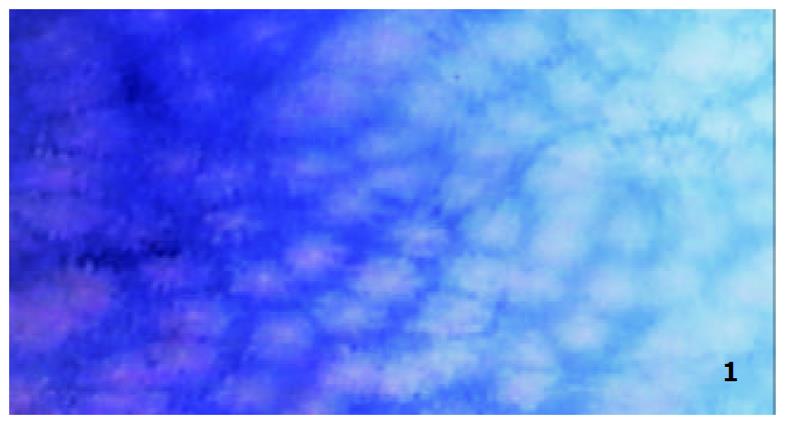
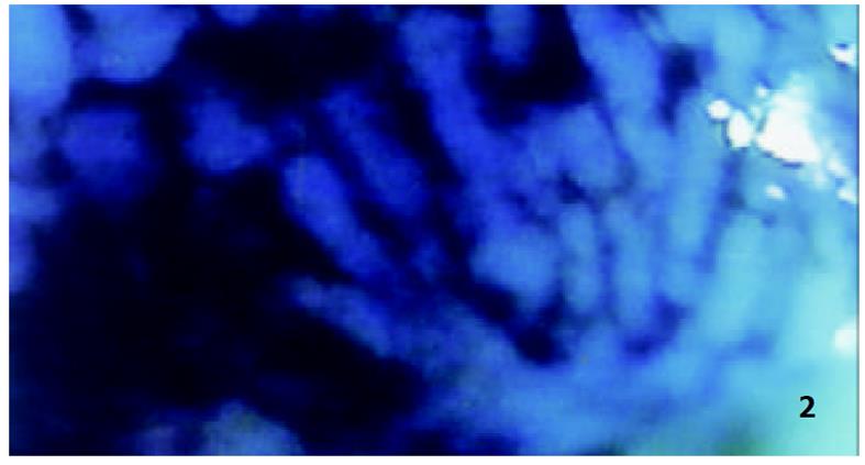
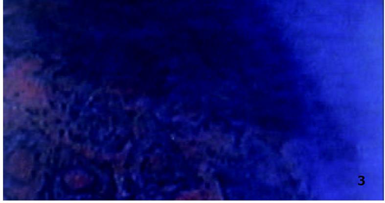
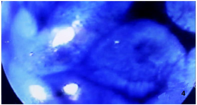
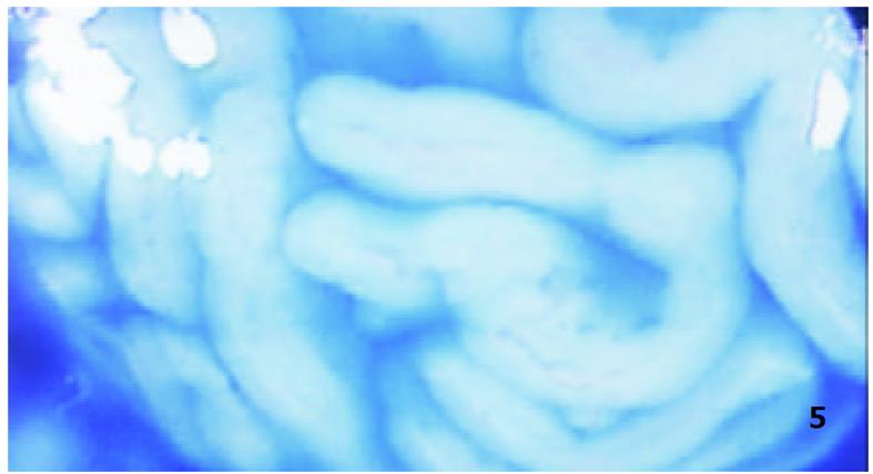
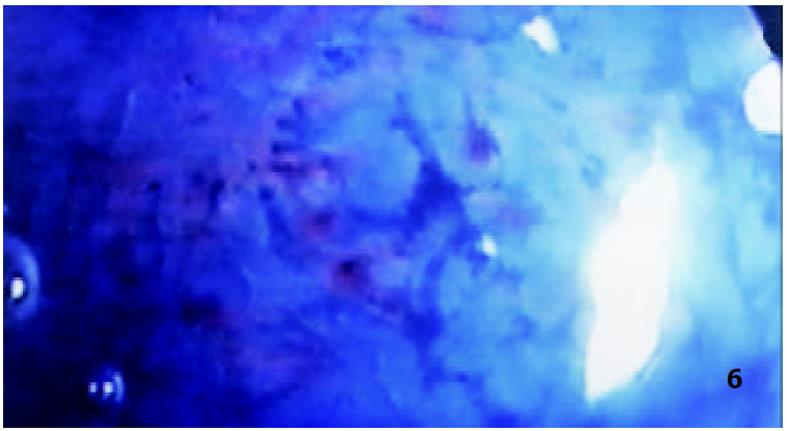
本组67例中单发息肉26例, 多发41例, 3例合并有结肠癌. 普通电子结肠镜发现息肉180颗, 经美蓝染色后用放大电子结肠镜检查又发现了1-3 mm大小的息肉90颗, 共计270颗. 美蓝染色后放大电子结肠镜检查明显提高了息肉的检出率(P<0.001, χ2 = 88.01)[10]. 息肉位于直肠31.1%; 乙状结肠24.4% ; 降结肠11.5% ; 横结肠24.8% ; 升结肠7.5%; 盲肠0.7%. 肉眼观察为山田I型息肉68.9%; 山田II型息肉18.1%; 山田III型息肉7.8%; 山田IV型息肉5.2%. 正常黏膜染色后用放大电子结肠镜观察隐窝开口呈均匀一致分布的圆形或类圆形结构(图1). 呈II型(图2)者2颗(0.7%); IIIS型(图3)者203颗(75.2%); IIIL型(图4)者49颗(18.1%); IV型(图5)者11颗(4.1%); Vn型(图6)共5颗(1.9%), 美蓝染色后电子放大结肠镜观察与组织病理学关系详见表1. 腺瘤性息肉大小与非典型增生及癌变的关系见表2. 13例腺瘤性息肉并非典型增生及癌变者, 于内镜电切术后1-3 mo行息肉残端染色放大电子结肠镜观察, 结果仅1例原为30 mm的息肉并中度非典型增生者, 其残端尚存10 mm大的腺瘤组织. 再次电切术, 术后未见再发. 其余患者病变残端均平坦, 美蓝染色后未见异常.
| 放大 分型 | 非瘤 n | 腺瘤性 息肉 n | 低度 非典型增生n | 中度 非典型增生n | 黏膜癌 n |
| II | 2 | ||||
| IIIS | 7 | 196 | |||
| IIIL | 42 | 5 | 2 | ||
| IV | 8 | 2 | 1 | ||
| Vn | 1 | 1 | 1 | 2 |
| 息肉最大径 | 非瘤 | 腺瘤 | 轻非典 | 中非典 | 癌变 |
| ≥2 mm | 1 (1.1) | 89 (88.9) | |||
| 3 mm- | 3 (2.6) | 116 (96.6) | 1 (0.8) | ||
| 6 mm- | 2 (5.0) | 35 (87.5) | 3 (7.5) | ||
| 10 mm- | 5 (71.4) | 2 (28.6) | |||
| 20 mm- | 1 (9.1) | 5 (45.5) | 2 (18.2) | 2 (18.2) | 1 (9.1) |
| 30 mm- | 1 (33.3) | 1 (33.3) | 1 (33.3) |
近来使用高分辨的电子结肠镜已使鉴别腺瘤性息肉成为可能[11]或将0.35 g/L靛蓝液加压喷洒到结肠息肉表面, 观察其渗血与否来判断是否为腺瘤[12], 其敏感性为92%, 大于普通结肠镜. 使用染色剂染色后再用放大结肠镜进行观察结肠黏膜隐窝形态对确定腺瘤性息肉更具有指导性意义[13]. 靛蓝胭脂染色是一种色素对比法, 他可显示出黏膜微细的凹凸变性, 再用放大结肠镜观察, 可较准确地预测腺瘤性息肉[14-19]、有无癌变[20,21]以及确定早期结肠癌浸润的深度, 对于制定治疗方法的选择也有指导意义. 美蓝染色剂是一种可吸收色素, 而腺管开口不染色, 这样可清楚显示腺管开口形态, 根据其形态变化帮助鉴别病灶的性质. 我们介绍的美蓝染色并经放大结肠镜观察后见正常结肠黏膜隐窝呈一致类圆形开口, 其他染色后类型也清晰地表现出来, 并且与组织学病理的结果相当一致. 我们检查的腺瘤性息肉中IIIS型占75.2%; IIIL型占18.1%, 其中伴有轻中度非典型增生者占14.3% (7/49); IV型占4.1%, 其中伴有非典型增生18.2%, 早期癌占9.1%; VN型占1.9%, 其中伴有中度非典型增生占20%, 早期癌占40%. 此结果与既往的报告类似. 即随着染色类型从III至V型, 则早期癌的检出率逐渐增多. 由于染色放大观察对癌变的腺瘤有较易识别的特性, 因此, 也可用于多发息肉特别是息肉病患者检查癌变息肉提供准确的方法. 染色后用放大结肠镜确定腺癌性息肉的准确率达到96.7%, 说明在进行病理检查前可预测其组织学类型. 对于发现癌变的腺瘤也有较高的价值. 由于我国腺瘤性息肉癌变的组织病理学诊断标准与日本相比存在较大差异. 因此, 早期癌的检出率低于日本文献报告的结果. 这也是今后进一步深入研究的方向. 本结果显示经美蓝染色放大电子结肠镜检查后明显提高了<3 mm的腺瘤性息肉的检出率, 这对于发现小的腺瘤并及早治疗起到了关键的作用.
| 9. | Kudo S, Rubio CA, Teixeira CR, Kashida H, Kogure E. Pit pattern in colorectal neoplasia: endoscopic magnifying view. Endoscopy. 2001;33:367-373. [PubMed] [DOI] |
| 11. | Eisen GM, Kim CY, Fleischer DE, Kozarek RA, Carr-Locke DL, Li TC, Gostout CJ, Heller SJ, Montgomery EA, Al-Kawas FH. High-resolution chromoendoscopy for classifying colonic polyps: a multicenter study. Gastrointest Endosc. 2002;55:687-694. [DOI] |
| 12. | Kanamori T, Itoh M, Yoshimi N. Pressuredye-spray: a simple and reliable method for differentiating adenomas from hyperplastic polyps in the colon. Gastrointest Endosc. 2000;55:695-700. [DOI] |
| 13. | Kudo S, Tamura S, Nakajima T, Yamano H, Kusaka H, Watanabe H. Diagnosis of colorectal tumorous lesions by magnifying endoscopy. Gastrointest Endosc. 1996;44:8-14. [DOI] |
| 14. | Kiesslich R, Von Bergh M, Hahn M, Hermann G, Jung M. Chromoendoscopy with indigocarmine improves the detection of adenomatous and nonadenomstous lesions in the colon. Endoscopy. 2001;33:1001-1006. [PubMed] [DOI] |
| 15. | Szaloki T. Indigo carmine contrast staining in combination with high resolution electronic endoscopy. Orv Hetil. 2002;143:25-29. [PubMed] |
| 16. | Togashi K, Konishi F, Ishizuka T, Sato T, Senba S, Kanazawa K. Efficacy of magnifying endoscopy in the differential diagnosis of neoplastic and non-neoplastic polyps of the large bowel. Dis Colon Rectum. 1999;42:1602-1608. [DOI] |
| 18. | Jaramillo E, Watanabe M, Slezak P, Rubio C. Flat neoplastic lesions of the colon and rectum detected by high-resolution video endoscopy and chromoscopy. Gastrointest Endosc. 1995;42:114-122. [DOI] |
| 19. | Matsumoto T, Iida M, Mizuno M, Shimizu M, Nakamura S, Fujishima M. In vivo observation of the ileal microadenoma in familia adenomatous polyposis. Am J Gastroenterol. 1999;94:3354-3358. [PubMed] [DOI] |
| 20. | Tung SY, Wu CS, Su MY. Magnifyipg colonoscopy in differentiating neoplastic from noneoplastic colorectal lesions. Am J Gastroenterol. 2001;96:2628-2632. [PubMed] [DOI] |
| 21. | Tamura S, Yokoyama Y, Tadokoro T, Higashidani Y, Kohsaki T, Onishi S. Depressed type submucosal invading colon cancer with type V pit pattern. Gastrointest Endosc. 2001;53:340-341. [DOI] |


