修回日期: 2003-01-20
接受日期: 2003-01-13
在线出版日期: 2003-08-15
探讨胸苷磷酸化酶(thymidine phosphorylase, TP)/血小板衍生内皮细胞生长因子(platelet-derived endothelial cell growth factor, PD-ECGF)和微血管密度(microvessel density, MVD)与大肠癌临床病理特征的关系.
应用免疫组化SP法, 检测50例大肠癌组织中胸苷磷酸化酶/即血小板衍生内皮细胞生长因子蛋白表达及微血管密度, 分析TP/PD-ECGF和MVD及其与大肠癌临床病理因素及预后的关系.
TP/PD-ECGF表达强度和MVD与大肠癌的肿块大小、Dukes分期、淋巴结转移、浸润深度密切相关 (P<0.01); 而与肿瘤分化无关(P>0.05). MVD与TP/PD-ECGF表达二者呈正相关(r = 0.72).
TP/PD-ECGF 与大肠癌血管生成密切相关, 对大肠癌的生长与浸润转移起促进作用. TP/PD-ECGF与MVD可作为反映大肠癌生物学行为的指标, 同时也是判断预后和指导辅助治疗的有效指标.
引文著录: 余细球, 邓长生, 朱尤庆, 程芳洲. 大肠癌组织胸苷磷酸化酶/血小板衍生内皮细胞生长因子的表达及意义. 世界华人消化杂志 2003; 11(8): 1197-1199
Revised: January 20, 2003
Accepted: January 13, 2003
Published online: August 15, 2003
To study the relationship between thymidine phosphorylase(TP)/platelet-derived endothelial cell growth factor(PD-ECGF), microvessel density (MVD) and the clinical pathological characteristics of colorectal carcinoma.
TP/PD-ECGF protein expression and microvessel density (MVD) in 50 colorectal carcinomas were examined by means of immunohistochemical staining S-P method and the relationship of TP/PD-ECGF, MVD and clinical pathological characteristics and its prognosis of colorectal cancers were analysed.
MVD and TP/PD-ECGF expression positively correlated with the size of the tumour, Dukes stage, lymph node metastasis and depth of invasion (P<0.01), but no significant correlation with the histologic type (P>0.05) was found.There was a positive correlation between MVD and TP/PD-ECGF expression (r = 0.72).
TP/PD-ECGF is highly related to angiogenesis of colorectal carcinoma and promotes invasion and metastasis. TP/PD-ECGF expression or MVD may be one of the predictors of the biological behaviors of colorectal carcinoma, and they may serve as prognostic factors and guide the treatment.
- Citation: Yu XQ, Deng CS, Zhu YQ, Cheng FZ. Expression and significance of thymidine phosphorylase/platelet-derived endothelial cell growth factor in human colorectal cancer tissue. Shijie Huaren Xiaohua Zazhi 2003; 11(8): 1197-1199
- URL: https://www.wjgnet.com/1009-3079/full/v11/i8/1197.htm
- DOI: https://dx.doi.org/10.11569/wcjd.v11.i8.1197
肿瘤的发生、发展与转移均需依赖新生血管的形成. 已知有许多影响血管生成的因子, 如胸苷磷酸化酶(thymidine phosphorylase, TP)即血小板衍化内皮细胞生长因子(platelet-derived endothelial cell growth factor, PD-ECGF)是该领域相对较新的一个血管因子. 为探讨大肠癌生物学行为中TP/PD-ECGF与肿瘤血管形成的作用, 应用免疫组化SP法检测人大肠癌组织中TP/PD-ECGF表达和微血管密度(microvessel density, MVD), 分析TP/PD-ECGF表达和MVD及其与大肠癌各临床病理指标间的关系.
我院1992/1995年手术切除并经病理证实的大肠癌石蜡标本50例, 男32例, 女18例, 年龄32-79 (平均58)岁. 术前均未进行过任何辅助治疗. Dukes: A期9例, B期10例, C期16例, D期15例; 发生淋巴结转移29例, 无淋巴结转移21例. 所有病例均获得术后5 a的随访资料.
制备4 μm连续切片3张, 1张用于HE染色, 另2张分别行免疫组化检测TP/PD-ECGF表达及MVD值(MVD检测用CD34标记内皮细胞). 采用免疫组化SP法, DAB显色. 微血管染色及TP/PD-ECGF染色用微波修复抗原. TP/PD-ECGF单克隆抗体、鼠抗人CD34 、SP试剂盒均购自北京中山生物试剂公司. TP/PD-ECGF表达阳性根据细胞染色强度和染色细胞所占面积二者积分来判断. 染色强度积分为: 不染色 = 0 ; 轻度染色 = 1; 中度染色 = 2; 强染色 = 3. 染色面积分为: 无细胞染色 = 0; <25%细胞染色 = 1; 25-50%细胞染色 = 2; >50%细胞染色 = 3. 若两种积分之和>2, 则为TP/PD-ECGF表达阳性, ≤2则为表达阴性[1]. 肿瘤微血管密度(MVD)测定先用低倍镜(×40倍)扫视整个切片, 寻找高血管密度区即"热点"区(即微血管生长集中区), 再转到高倍镜(×200倍)下精确计数微血管数量. 取3个热点区微血管数量的平均值视作为每例的微血管数[2].
统计学处理 TP/PD-ECGF以(+)或(-)表示, 为计数资料; MVD以(mean±SD)表示. TP/PD-ECGF和MVD之间关系用t检验; TP/PD-ECGF与临床病理因素之间用χ2检验; MVD与临床病理因素之间关系用t检验, 与肿瘤分期关系用单因素方差分析和t检验; 生存率采用Kaplan-Meier法计算, 生存期差别的比较用Log-rank 检验; P<0.05, 有统计学意义.
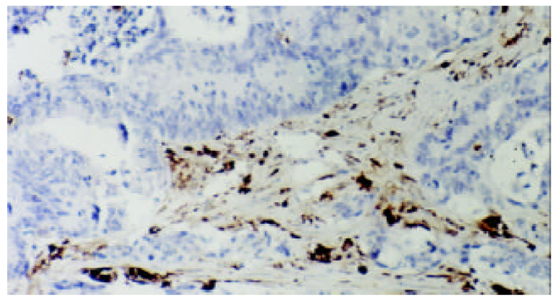
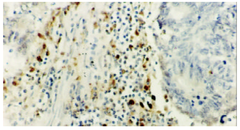
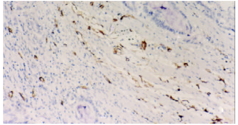
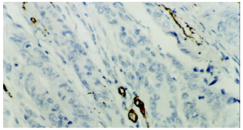
在50例标本中, TP/PD-ECGF阳性35例, 阳性率70%. TP/PD-ECGF阳性染色主要定位于肿瘤间质细胞胞质, 呈棕黄色. 偶有胞核染色. 而肿瘤细胞团阳性染色少. CD34+将微血管内皮细胞胞质染成黄褐色, 呈环状. 微血管密集于肿瘤基质, 尤其肿瘤浸润前缘的微血管较肿瘤内部密集, MVD值10-57, 平均为36.5±10.2 (图1, 图2, 图3, 图4).
TP/PD-ECGF阳性组中的MVD值45.5±10.2明显高于TP/PD-ECGF阴性组20.3±9.3 (P<0.01). TP/PD-ECGF表达与肿块大小(P<0.05)、Dukes分期(P<0.05)、浸润深度(P<0.01)、淋巴结转移(P<0.01)密切相关, 与肿瘤分化程度无关(P>0.05). MVD值在肿瘤大于5 cm 组明显高于小于5 cm组(P<0.05), 肿瘤侵及浆膜外组明显高于肿瘤局限于浆膜组 (P<0.01), 有淋巴结转移组MVD高于无淋巴结转移组(P<0.01), MVD与肿瘤分期关系显示各期的MVD值有差别 (P<0.05), 提示随着肿瘤分期增加而增加, 但与肿瘤分化程度无关 (表1) .
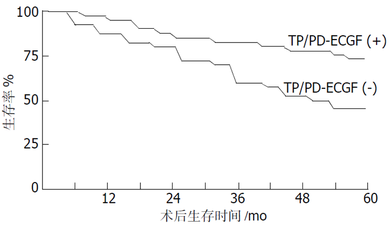
在50例患者中, 有26例术后存活5 a或5 a以上, 5 a存活率为52%. 其中TP阳性组术后5 a存活率42.8%, 明显低于TP阴性组的术后5 a生存率73.3%, (P<0.01, 图5).
胸苷磷酸化酶(TP)是嘧啶合成分解过程中的一个酶, 与血小板衍化内皮细胞生长因子(PD-ECGF)为同一物质. 他能可逆催化胸腺嘧啶核苷为胸腺嘧啶和2-脱氧-核糖-1-磷酸, 后者去磷酸化的产物2-脱氧-D-核糖具有血管生成和催化内皮细胞活性[3]. 该酶在体外具内皮细胞催化性, 体内有血管生成活性, 是少有的具酶活性的促血管生长因子, 主要分布于人体血小板和胎盘, 他在宫颈癌、肝癌、胃癌、大肠癌等许多实体瘤中异常增高, 其表达增高往往预示其预后不良[4-6]. TP/PD-ECGF与宫颈癌、食管癌、胃癌、肝癌组织中MVD密切相关, 甚至可使肿瘤细胞凋亡减少[7-10]. TP/PD-ECGF、MVD一起成为许多实体瘤的发生、发展及预后的评判指标.
我们发现在大肠癌中TP/PD-ECGF阳性组其MVD高, 其表达强度与MVD值呈正相关. 从免疫组化图片上, 我们发现TP/PD-ECGF 阳性表达多见于肿瘤基质也即微血管丰富区, 而少微血管的肿瘤细胞团也少有TP/PD-ECGF阳性表达. 说明TP/PD-ECGF与肿瘤微血管密度密切相关, 位于肿瘤间质的TP/PD-ECGF对大肠癌肿瘤的微血管生成与发展起重要作用. 这与Tanioka et al [11]在脑星形细胞瘤中观察到的结果一致. 同时还发现TP/PD-ECGF表达强度也与肿块大小、浸润深度、分期及淋巴结转移密切相关. TP/PD-ECGF阳性表达多见于肿块大于5 cm、肿瘤浸润深及浆膜外、分期高、伴淋巴结转移的大肠癌患者. 表明TP/PD-ECGF作为促血管生长因子, 是大肠癌血管形成与维持的重要因子, 对大肠癌的生长、浸润、转移起重要作用.
我们还发现, TP/PD-ECGF表达、MVD与大肠癌预后也有相关性. TP/PD-ECGF阳性组其MVD值(46±10)明显高于TP/PD-ECGF阴性组MVD值(20±13), 且TP/PD-ECGF阳性组其术后5 a生存率42.8%明显低于TP/PD-ECGF阴性组73.3%. 因此, 检测大肠癌中TP/PD-ECGF不仅可以反映大肠癌内微血管生成情况, 也可以反映大肠癌生长、浸润、转移能力, 同时也反映其预后情况.
5-氟尿嘧啶(5-FU)是临床上广泛用于结肠癌、乳腺癌等肿瘤化疗药. TP/PD-ECGF酶能催化5-FU与及其前体物质(如: 氟铁龙、希罗达capecitabine), 阻断DNA合成, 消除潜在血管生长因子[12,13]. 实现抗肿瘤生长与抗肿瘤血管生成的统一. 因TP/PD-ECGF表达在肿瘤组织中高于正常组织, 故在TP/PD-ECGF酶表达增高的肿瘤如消化道肿瘤、乳腺癌等, 应用5-FU及其前体药具有确切疗效. 因此检测肿瘤组织中TP/PD-ECGF表达也是提示化疗有效的一个预测因素, 具有重要的临床意义.
| 1. | Mattern J, Koomagi R, Volm M. Biological characterization of subgroups of squamous cell lung carcinomas. Clin Cancer Res. 1999;5:1459-1463. [PubMed] |
| 2. | Weidner N. Current pathologic methods for measuring intratumoral microvessel density within breast carcinoma and other solid tumors. Breasr Cancer Res Treat. 1995;36:169-180. [DOI] |
| 3. | Stevenson DP, Milligan SR, Collins WP. Effects of platelet-derived endothelial cell growth factor/thymidine phosphorylase, substrate, and products in a three-dimensional model of angiogenesis. Am J Pathol. 1998;152:1641-1646. [PubMed] |
| 4. | Takebayashi Y, Yamada K, Miyadera K, Sumizawa T, Furukawa T, Kinoshita F, Aoki D, Okumura H, Yamada Y, Akiyama S. The activity and expression of thymidine phosphorylase in human solid tumours. Eur J Cancer. 1996;32:1227-1232. [DOI] |
| 5. | Jin-no K, Tanimizu M, Hyodo I, Nishikawa Y, Hosokawa Y, Endo H, Doi T, Mandai K, Ishitsuka H. Circulating platelet-derived endothelial cell growth factor increases in hepatocellular carcinoma patients. Cancer. 1998;82:1260-1267. [DOI] |
| 6. | Fujimoto J, Sakaguchi H, Hirose R, Wen H, Tamaya T. Clinical implication of expression of platelet-derived endothelial cell growth factor(PD-ECGF) in metastatic lesions of uterine cervical cancers. Cancer Res. 1999;59:3041-3044. [PubMed] |
| 7. | Tang W, Wang X, Utsunomiya H, Nakamuta Y, Yang Q, Zhang Q, Zhou G, Tsubota Y, Mabuchi Y, Li L. Thymidine phosphorylase expression in tumor stroma of uterine cervical carcinomas: histological features and microvessel density. Cancer Lett. 2000;148:153-159. [DOI] |
| 8. | Okamoto E, Osaki M, Kase S, Adachi H, Kaibara N, Ito H. Thymidine phosphorylase expression causes both the increase of intratumoral microvessel and decrease of apoptosis in human esophageal carcinomas. Pathol Int. 2001;51:158-164. [PubMed] [DOI] |
| 9. | Osaki M, Sakatani T, Okamoto E, Goto E, Adachi H, Ito H. Thymidine phosphorylase expression results in a decrease in apoptosis and increase in intratumoral microvessel density in human gasrtric carcinomas. Virchow Arch. 2000;437:31-36. [DOI] |
| 10. | Guo L, Kuroda N, Toi M, Miyazaki E, Hayashi Y, Enzan H, Jin Y. Increased expression of platelet-derived endothelial cell growth factor in human hepatocellular carcinomas correlated with high Edmondson grades and portal vein tumor thrombosis. Oncol Rep. 2001;8:871-876. [DOI] |
| 11. | Tanioka K, Takeshima H, Hirano H, Kimura T, Nagata S, Akiyama S, Kuratsu J. Biological role of thymidine phosphorylase in human astrocytic tumors. Oncol Rep. 2001;8:491-496. [DOI] |
| 12. | Griffiths L, Stratford J. Platelet-derived endothelial cell growth factor/thymidine phosphorylase in tumor growth and response to therapy. British J Cancer. 1997;76:689-693. [DOI] |


