Copyright
©The Author(s) 2023.
World J Gastroenterol. Jul 14, 2023; 29(26): 4200-4213
Published online Jul 14, 2023. doi: 10.3748/wjg.v29.i26.4200
Published online Jul 14, 2023. doi: 10.3748/wjg.v29.i26.4200
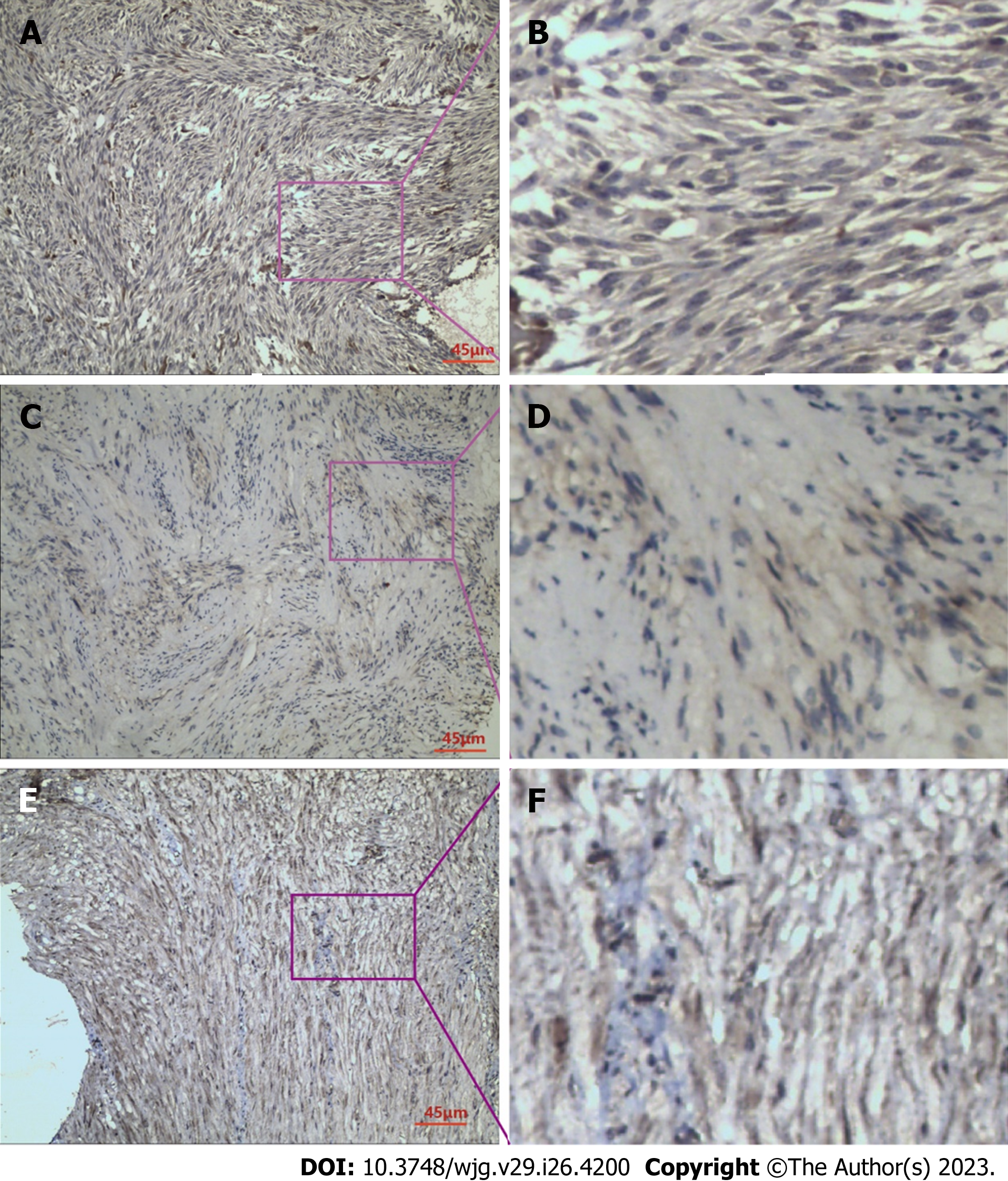
Figure 1 Immunohistochemical staining.
A: Immunohistochemical staining for raf kinase inhibitor protein (RKIP) in gastrointestinal stromal tumor (GIST) tissues; B: RKIP protein showed positive expression in the cytoplasm and cell membrane, appearing as brownish-yellow or brown granules; C: Immunohistochemical staining for phosphorylated-extracellular signal-regulated kinase (P-ERK) in GIST tissues; D: P-ERK protein exhibited heterogeneous distribution in GIST cells, mainly in the cytoplasm, with occasional presence in the nucleus, and appeared as brownish-yellow granules; E: Immunohistochemical staining for phosphorylated-mitogen-activated protein kinase/ERK Kinase (P-MEK) in GIST tissues; F: P-MEK protein expression in GIST cells was observed as brownish-yellow granules, mainly distributed in the cytoplasm, with some in the nucleus, and showed a relatively uniform distribution. A, C and E were taken under 200 × magnification.
- Citation: Qu WZ, Wang L, Chen JJ, Wang Y. Raf kinase inhibitor protein combined with phosphorylated extracellular signal-regulated kinase offers valuable prognosis in gastrointestinal stromal tumor. World J Gastroenterol 2023; 29(26): 4200-4213
- URL: https://www.wjgnet.com/1007-9327/full/v29/i26/4200.htm
- DOI: https://dx.doi.org/10.3748/wjg.v29.i26.4200


