Copyright
©The Author(s) 2023.
World J Gastroenterol. Mar 21, 2023; 29(11): 1669-1684
Published online Mar 21, 2023. doi: 10.3748/wjg.v29.i11.1669
Published online Mar 21, 2023. doi: 10.3748/wjg.v29.i11.1669
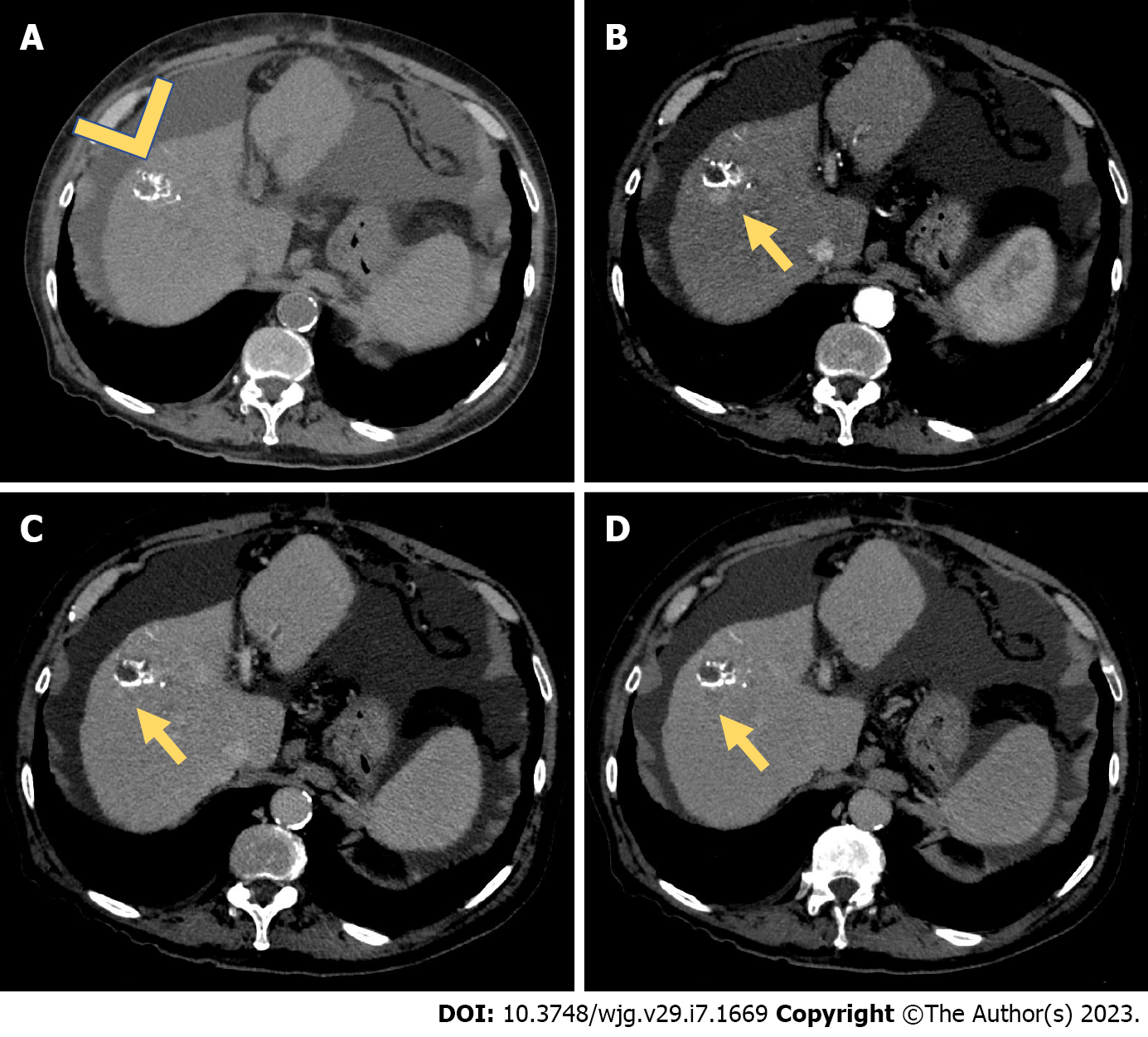
Figure 2 Computed tomography study for assessment of treatment response (after transarterial chemoembolization).
A: A 55-year-old male underwent conventional transarterial chemoembolization of a hepatocellular carcinoma lesion located in the VIII hepatic segment. Two years after treatment, a computed tomography scan showed areas of hyperattenuating components in the unenhanced phase (A), representing the ethiodized oil (arrowhead); B-D: During the arterial (B) phase, a pseudonodular area of hypervascularization during the arterial phase (B, yellow arrow) was seen, with a slight hypoattenuating appearance during the portal venous phase (C) and a clear washout during the delayed phase (D). This represents an example of recurrent hepatocellular carcinoma after transarterial chemoembolization.
- Citation: Ippolito D, Maino C, Gatti M, Marra P, Faletti R, Cortese F, Inchingolo R, Sironi S. Radiological findings in non-surgical recurrent hepatocellular carcinoma: From locoregional treatments to immunotherapy. World J Gastroenterol 2023; 29(11): 1669-1684
- URL: https://www.wjgnet.com/1007-9327/full/v29/i11/1669.htm
- DOI: https://dx.doi.org/10.3748/wjg.v29.i11.1669


