Copyright
©The Author(s) 2022.
World J Gastroenterol. Feb 21, 2022; 28(7): 755-762
Published online Feb 21, 2022. doi: 10.3748/wjg.v28.i7.755
Published online Feb 21, 2022. doi: 10.3748/wjg.v28.i7.755
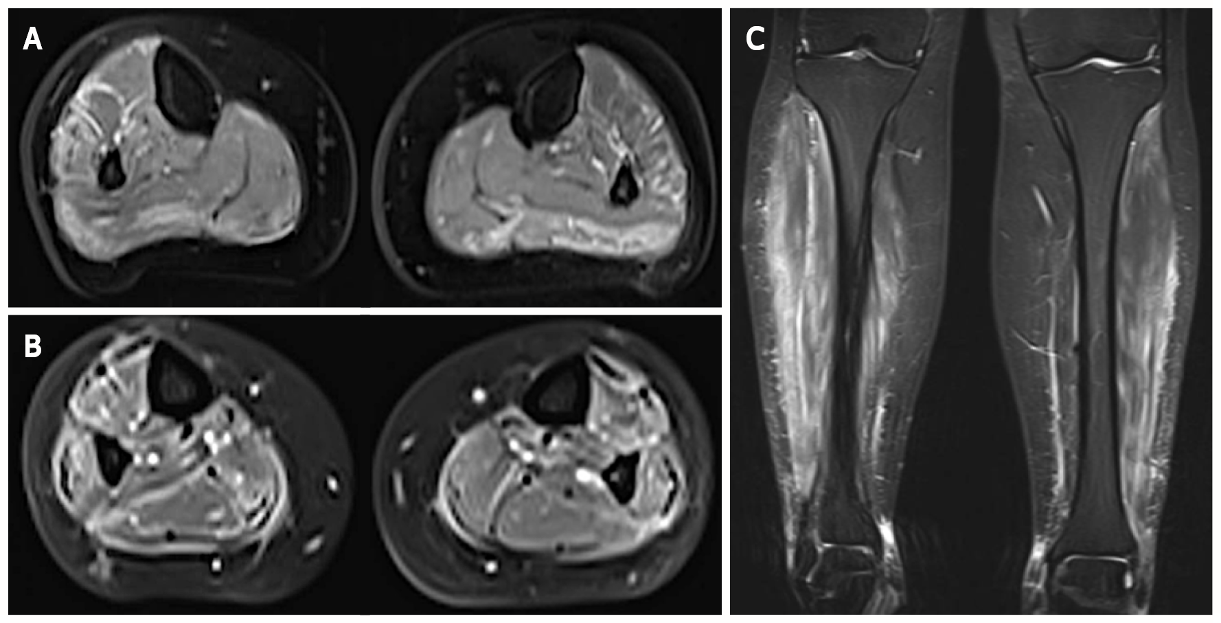
Figure 1 Magnetic resonance imaging of the calves in axial and frontal views.
A and C: Demonstration of high signal on T2-weighted images in muscles from posterior and lateral compartments of the legs as well as in their surrounding fascia; B: This finding was accentuated on gadolinium-enhanced fat-suppressed T1-weighted images, suggestive of myositis and fasciitis.
- Citation: Catherine J, Kadhim H, Lambot F, Liefferinckx C, Meurant V, Otero Sanchez L. Crohn’s disease-related ‘gastrocnemius myalgia syndrome’ successfully treated with infliximab: A case report. World J Gastroenterol 2022; 28(7): 755-762
- URL: https://www.wjgnet.com/1007-9327/full/v28/i7/755.htm
- DOI: https://dx.doi.org/10.3748/wjg.v28.i7.755


