Copyright
©The Author(s) 2020.
World J Gastroenterol. Jun 14, 2020; 26(22): 2967-2986
Published online Jun 14, 2020. doi: 10.3748/wjg.v26.i22.2967
Published online Jun 14, 2020. doi: 10.3748/wjg.v26.i22.2967
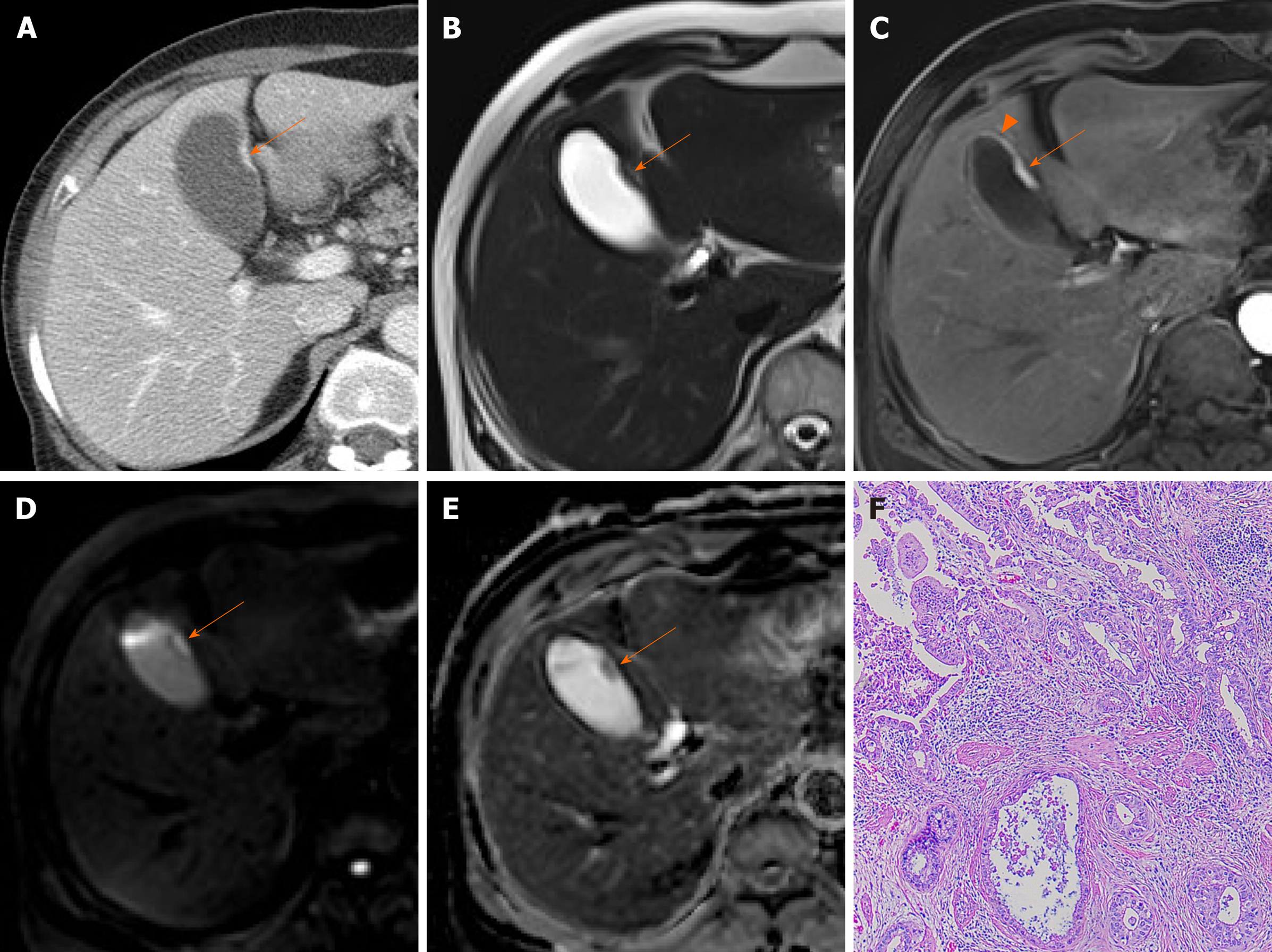
Figure 9 Cancer of gallbladder body.
A: Flat, thickened wall (arrow) with single enhancing layer of gallbladder body on computed tomography; B: No observable intramural cysts on T2-weighted image; C: Strongly enhanced lesion (arterial phase), with mild thickening of adjacent fundal wall (arrowhead) on magnetic resonance imaging; D, E: Obvious diffusion restriction on diffusion-weighted imaging (D) and apparent diffusion coefficient map (E); F: Adenocarcinoma confirmed on hematoxylin and eosin staining (×100) after cholecystectomy.
- Citation: Yu MH, Kim YJ, Park HS, Jung SI. Benign gallbladder diseases: Imaging techniques and tips for differentiating with malignant gallbladder diseases. World J Gastroenterol 2020; 26(22): 2967-2986
- URL: https://www.wjgnet.com/1007-9327/full/v26/i22/2967.htm
- DOI: https://dx.doi.org/10.3748/wjg.v26.i22.2967


