Copyright
©The Author(s) 2017.
World J Gastroenterol. Dec 28, 2017; 23(48): 8671-8678
Published online Dec 28, 2017. doi: 10.3748/wjg.v23.i48.8671
Published online Dec 28, 2017. doi: 10.3748/wjg.v23.i48.8671
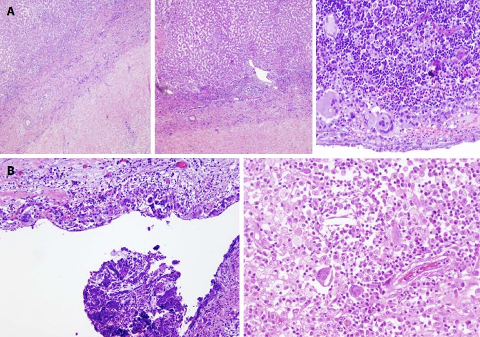
Figure 4 Histological examination of the surgical specimens from the three cases of xanthogranulomatous cholecystitis reveals findings of chronic cholecystitis and marked inflammatory infiltrate, including lymphocytes, plasma cells, foamy histiocytes, and spindle-shaped cells.
A: Focal formations of pseudocysts, with multinucleated foreign-body giant cells and cholesterol clefts, are also observed; B: Hyalinization and fibrosis of the gallbladder wall reflects chronic inflammation. Typically, the xanthogranulomatous reaction occupies a limited portion of the gallbladder wall, while the remainder shows signs of conventional chronic cholecystitis. Polymorphonuclear lymphocytes, reflecting acute inflammation, are also occasionally seen. The mucosa presents focal ulceration and erosion and reactive changes that consist in papillary hyperplasia and mucinous and cardial-type glandular metaplasia. Dysplastic changes and malignant features are absent in all three cases.
- Citation: Nacif LS, Hessheimer AJ, Rodríguez Gómez S, Montironi C, Fondevila C. Infiltrative xanthogranulomatous cholecystitis mimicking aggressive gallbladder carcinoma: A diagnostic and therapeutic dilemma. World J Gastroenterol 2017; 23(48): 8671-8678
- URL: https://www.wjgnet.com/1007-9327/full/v23/i48/8671.htm
- DOI: https://dx.doi.org/10.3748/wjg.v23.i48.8671


