Copyright
©The Author(s) 2017.
World J Gastroenterol. Oct 14, 2017; 23(38): 7047-7053
Published online Oct 14, 2017. doi: 10.3748/wjg.v23.i38.7047
Published online Oct 14, 2017. doi: 10.3748/wjg.v23.i38.7047
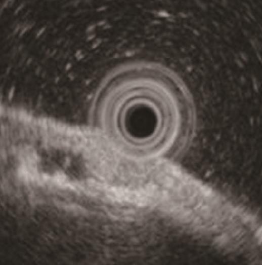
Figure 3 Endoscopic ultrasonography findings.
The tumor slightly invaded the third layer, although it was located mainly in the second layer. Moreover, a hypoechoic mass located in the third layer near the tumor was detected.
- Citation: Manabe S, Mukaisho KI, Yasuoka T, Usui F, Matsuyama T, Hirata I, Boku Y, Takahashi S. Gastric adenocarcinoma of fundic gland type spreading to heterotopic gastric glands. World J Gastroenterol 2017; 23(38): 7047-7053
- URL: https://www.wjgnet.com/1007-9327/full/v23/i38/7047.htm
- DOI: https://dx.doi.org/10.3748/wjg.v23.i38.7047


